Share this project
Paweena Diloksumpan
Universiteit Utrecht
Publication date: 7 juli 2020
University: Universiteit Utrecht
ISBN: 978-94-6380-852-1
The Use of Converging Biofabrication Techniques to Address Nature’s Complexity in Joint Repair
Summary
Three-dimensional (3D) printing technologies offer multiple possibilities for the custom-tailored fabrication of either cartilage or bone grafts. However, employing this technology for the integration of different materials that possess mechanically distinct properties, is still a major challenge in tissue engineering and regenerative medicine. To address this challenge, two main approaches were developed in this thesis: 1) the development of printable materials for use as a bone scaffold with physical and chemical properties that permit their patterning in direct contact with the microfiber-reinforced hydrogels that are used as chondral substitutes, and 2) the development of a multi-scale printing process for integrating such bone and cartilage compartments. Therefore, this thesis presents a promising strategy for combining different 3D printing technologies to integrate microfiber-reinforced, hydrogel-based chondral constructs and calcium phosphate-based subchondral bone compartments, in order to establish and engineer a functional osteochondral interface.
Recently, the compressive properties of gelatin methacrylate hydrogels (gelMA) were increased to values comparable to those of native cartilage, by reinforcing cast gels with polycaprolactone (PCL) microfiber meshes that were produced by melt electrowriting (MEW), a microscale 3D printing technology. To further improve such composite structures and enable their application in large osteochondral defects, the anchoring of such reinforced structures on osteal scaffolds in a way that mimics the natural subchondral bone, was addressed in this thesis (Chapter 3). Highly organized microfiber lattices produced by MEW using PCL, a thermoplastic synthetic polymer susceptible to melt at relatively low temperatures (approximately 60°C), were shown to preserve their unique architecture (responsible for the mechanical reinforcement that is observed in combination with infused hydrogels), when combined with low-temperature setting ceramics.
For this purpose, a calcium phosphate-based material that could set at physiological temperature, with alpha-tricalcium phosphate (α-TCP) as main ceramic component, was developed and combined with a custom-synthesized thermo-reversible hydrogel carrier. Such composite bioceramics can be solidified at low temperature, forming calcium-deficient hydroxyapatite (CDHA). This process is compatible with the inclusion of labile materials like PCL. The mixture of α-TCP with the thermo-reversible hydrogel endowed the system with shear-thinning properties that allowed the use of the composite as bio-ink for printing with an extrusion-based printer at ambient temperature, hence facilitating the fabrication of a subchondral bone plate-mimicking interface and a cancellous bone-mimicking structure underneath. These integrated PCL-CDHA structures were obtained after solidification of directly dispensed printable calcium phosphate-based composite (pCaP) onto the PCL microfiber reinforcing framework of the chondral compartment. The adequate and secure integration permitted to greatly increase the shear strength of the engineered bone-to-cartilage boundary, which permitted additional manipulation of the construct, such as hydrogel infusion in the chondral region and implantation in surgically produced osteochondral defects ex vivo and in vivo.
Prior to the in vivo translation of these composite biomaterials, this thesis extensively investigated the mechanical and biological performance in vitro of the constructs, thereby focusing on unravelling the bone-forming potential of the osteal anchor, a necessary step for functional healing of osteochondral defects. The porous CDHA structures produced by 3D printing appeared to exhibit compressive properties within the range of human cancellous bone when porosity ranged between 20% and 60%. Additionally, bone-marrow derived mesenchymal stromal cells were shown to proliferate both within CDHA-incubated medium and on the printed CDHA itself, and to differentiate toward the osteogenic lineage when supplemented with osteogenic induction factors. These positive findings justified proceeding with in vivo evaluations. For that purpose, cylindrical, 3D printed bony scaffolds displaying different pore distributions, with either a constant porosity throughout the scaffold or a decreasing gradient of porosity along the axial direction of the cylinder (as in natural subchondral bone), were tested in a large (cm-scale) critical size bone defect in a non-load-bearing orthotopic location in an equine model (Chapter 4). A PCL case around the cylinder permitted invasion of the newly-formed bone only from the bottom. Results demonstrated better scaffold resorption, paired with more and more homogeneous bone regeneration in the implants with constant porosity. Given this superior performance, the constant porosity architecture was chosen for the further development of the osteochondral construct for cartilage repair at a load-bearing site.
The final concepts of the osteochondral graft were tested in two in vivo studies in ponies in which the chondral compartments had a different composition and architecture. In both implants the bone phase and interphase between the bone and the cartilage phase were similar consisting of the pCaP composite with anchored MEW fibers as osteal anchor and subchondral bone phase of the graft. In the first study, the chondral phase was composed by articular cartilage derived progenitor cells (ACPCs) seeded within the PCL mesh, and stimulated during pre-culture in vitro with growth differentiation factor-2 (GDF-2), resulting in the formation of a cartilaginous disc entangled within the polymeric microfibers (Chapter 5). In the second study, ACPC-laden gelMA was co-printed with the PCL meshwork and the constructs were cultured for a month with chondrogenic differentiation media (Chapter 6). Cell-free controls were used in both studies. In all cases, regardless of the configuration of the implant, the scaffolds permitted ACPCs to proliferate and produce cartilage-related extracellular matrix molecules, such as glycosaminoglycans (GAGs) and type II collagen during the 4-week culture prior to implantation. After an implantation for 6 months, the results were totally different for the two studies, which seemed to be due to striking differences in the performance of the bony phases.
In the GDF-2 study, at 6 months after implantation the GAG-content of the cell-laden structure had substantially decreased compared to the level after pre-culture. There was also a minimal volume of newly-formed bone, and a rather heavy inflammatory reaction was observed after implantation for 6 months, both in the cell-laden group and in the cell-free control. Additionally, misalignment and misposition of the grafts was clearly visible.
In the other group, pre-culture GAG content had slightly increased from the time of implantation until 6 months after implantation in the study. Interestingly, a similar GAG content was observed in cell-free controls. The volume of newly-formed bone in the subchondral bone compartment was higher compared with the GDF-2 study and there was much less misalignment.
It was hypothesized that the imperfect geometry of the printed subchondral bone compartment in the GDF-2 study, combined with the brittleness of the ceramic material might have led to structural damage of the implants during surgical implantation, fragilizing the structure and eventually leading to its collapse when subjected to loading, and overall implant failure. This had not happened in the other study thanks to the use of a mold in that study when producing the bone phase, which led to better shape fidelity post-printing and thus better geometrical fitting in the defects. These studies, while showing the appropriateness of the concept and the feasibility of the proposed approach, also clearly demonstrated the limitations of the current, brittle, pCaP composite for application at load-bearing sites. Finally, the equine model showed to be a challenging, but relevant model as a translational model preparing for clinical use in human and animal patient populations.
It can be concluded that the work in this thesis has produced substantial progress towards the final goal of clinical translation of fully mechanically competent and regenerative osteochondral grafts by the clever use of various 3D-printing technologies to integrate two mechanically distinct materials together to form an entire osteochondral graft. Nevertheless, there are several challenges that still need to be explored further, especially regarding the development of alternative materials with higher fracture toughness for the bone compartment. Additionally, improvements can be made to the interface itself as well. For instance, with respect to permeability and its role in load distribution and dissipation. Better recapitulation of the natural situation may be the way to go here.
List of Abbreviations
2D two-dimensional
3D three-dimensional
ACPCs articular cartilage-derived progenitor cells
ALP alkaline phosphatase
AM additive manufacturing
APS ammonium persulphate
ASAP L-ascorbic acid 2-phosphate
α-MEM minimum essential medium alpha
α-TCP alpha-tricalcium phosphate
bFGF basic fibroblast growth factor
BID twice a day
BMP-7 bone morphogenetic factor-7
β-GP beta-glycerophosphate
C crosslinkable
CAD computer aided design
CaP calcium phosphate
CBC complete blood count
CDHA calcium deficient hydroxyapatite
C-pCaP crosslinkable printable calcium phosphate
CR ceramic remnant
DAB 3, 3-diaminobenzidine-horseradish peroxidase
DLP digital light projection
DMA dynamic mechanical analyzer
DMMB dimethylmethylene blue
ECM extracellular matrix
FCS fetal calf serum
FDM fused deposition modelling
GAGs glycosaminoglycans
GDF-2 growth differentiation factor-2
gelMA methacryloyl-modified gelatin
GRF ground reaction force
HEPES N-2-hydroxyethylpiperazine-N-2-ethane sulfonic acid
H&E hematoxylin and eosin
IGF-1 insulin-like growth factor-1
IM intramuscular
IV intravenous
LDH lactate dehydrogenase
LVR linear viscoelastic range
MEW melt electrowriting
MMA methyl methacrylate
MRI magnetic resonance imaging
MSCs mesenchymal stem cells, mesenchymal stromal cells
µ-CT micro-computed tomography
NaOH sodium hydroxide
Nano-HA nano-hydroxyapatite
NB new bone ingrowth
NC non-crosslinkable
NC-pCaP non-crosslinkable printable calcium phosphate
OC osteochondral
PCL polycaprolactone
PDMS polydimethylsiloxane
PLA polylactic acid
PO oral administration
ROI region of interest
ROM range of motion
SD standard deviation
SEM scanning electron microscopy
SID once a day
TEMED tetramethylethylenediamine
TGF-β3 transforming growth factor b3
TRAP tartrate-resistant acid phosphatase
VOI volume of interest
XRD x-ray diffraction pattern
See also these dissertations

Toby van Gastelen
Structure-Preserving Data-Driven Methods for Modeling Turbulent Flows
19 mei 2026
TU Eindhoven
Open Ebook
Ton Winkelmolen
Molecular insights into the role of VRS5 in tillering and lateral spikelet development in barley
12 juni 2026
Wageningen University
Open Ebook
Selcuk Peker
Gamma Knife Radiosurgery for Skull Base Tumors
10 juni 2026
Universiteit Maastricht
Open Ebook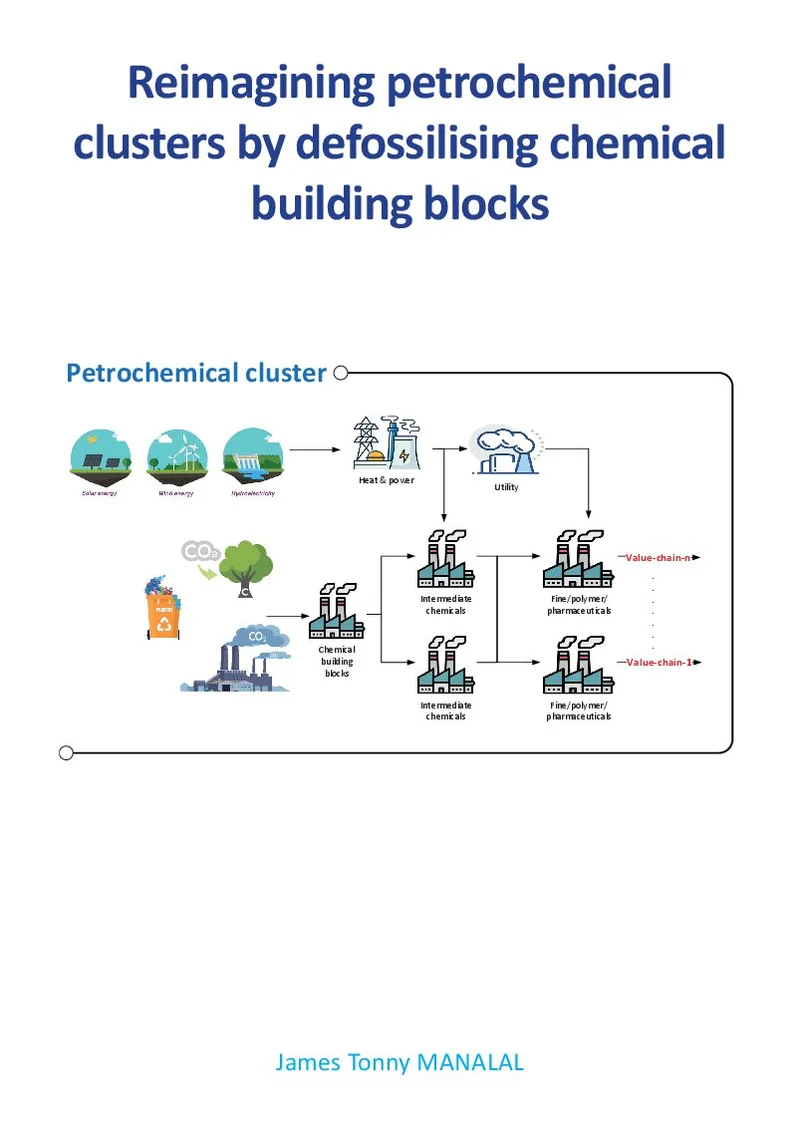
Tonny Manalal
Reimagining petrochemical clusters by defossilising chemical building blocks
18 mei 2026
Overig
Open Ebook
Cora De Gol
Microbial stabilization and protein functionality of plant-based liquids using pulsed electric fields
17 juni 2026
Wageningen University
Open Ebook
We print for the following universities


