Share this project
Eric Alexander Dik
Universiteit Utrecht
Publication date: 15 oktober 2019
University: Universiteit Utrecht
ISBN: 978-94-6380-524-7
Early Stage Oral Squamous Cell Carcinoma
Summary
Conclusions chapter 2
-The histological growth parameters PG, VG and IG determined on the resection specimen could be related to the presence of occult metastasis. These parameters have in consequence prognostic value.
-There is a poor correlation between histological parameters determined on the biopsy and resection specimen. Especially biopsies that lack a specific histological parameter are not reliable and can reassure the clinician wrongly.
-Reliable determination of tumor infiltration depth is not possible in the majority (85%) of cases because the biopsy thickness does not comprise the tumor thickness.
-Increase of the biopsy diameter by ≥ half of the tumor diameter, could improve the predictive value of the biopsy.
-In this group of patients, the biopsy does currently not add any value in the treatment planning of the cN0 neck.
In case of an OSCC a distinction is made between well, moderately, poorly and undifferentiated tumors. In many head and neck cancer centers the biopsy specimen as well as the resection specimen is routinely graded. Deterioration of grade is related to aggressive tumor behavior and metastasis. This relation is controversial however. In chapter 3 the value of the biopsy as a predictor for the differentiation grade in the subsequent resection specimen was analyzed. To analyze if grading of OSCC’s on the biopsy and resection specimen is of added value the following topics were covered:
1. The relation between differentiation grade, determined on the biopsy- and resection specimen, and the occurrence of occult metastasis and survival was determined.
2. The predictive value of the biopsy was determined by analyzing the correlation between biopsy and resection specimen concerning the determination of differentiation grade.
3. Deterioration of grade determined on the resection specimen was related to the presence of PG, VG and IG.
4. The effect of adding IG to the differentiation grade on the correlation with the N stadium, was analyzed.
In total 145 patients with a pT1-2 cN0 OSCC of the tongue, floor of mouth or cheek were included.
-Deterioration of differentiation grade on the biopsy- as well as the resection specimen was not related to the presence of occult metastasis (resp. p=1.0 and p=0.50) nor to survival (resp. p=0.65 and p=0.44).
-Correlation between biopsy and subsequent resection specimen concerning differentiation grade is poor. A similar differentiation grade in biopsy- and resection specimen considering well, moderately and poorly differentiated tumors, was found in resp. 43%, 83% and 39% of the cases.
-Only in case of poorly differentiated tumors, a significantly higher amount of VG was found (p=0.02). No significant relation could be found between the presence of the unfavorable histological parameters PG and IG and the deterioration of differentiation grade (resp. p=0.15 and p=0.85).
-Only the combination of IG with a moderately differentiation grade gave a significantly higher risk on nodal metastasis (p=0.02). Combining the pattern of invasion with well and poorly OSCC’s did not lead to a significantly higher risk on nodal metastasis (resp. p=0.62 and p=0.47).
Conclusions chapter 3
-Concerning the poor correlation of differentiation grade between biopsy and resection specimen, the differentiation grade determined on the biopsy must be considered as a poor predictor for the subsequent resection specimen.
-Since deterioration of the differentiation grade could not be related to the presence of nodal metastasis nor survival, these criteria don’t have any prognostic value concerning outcome and are consequently of little value for treatment planning.
-By adding the histological parameter IG to the differentiation grade, the prognostic value could increase, probably because it is an independent risk factor for nodal metastasis.
The pathology report
In case of an OSCC the pathology report is a dictating tool whether to perform an adjuvant therapy after primary surgery. Pathologists assess more and more on digital slides. Since the pathology report is a determining factor, knowledge about the inter-observer variation (IOV) is relevant to estimate the level of reliability and reproducibility of the report. In chapter 4 the pathology report was highlighted. The focus was on the IOV during the determination of the histological parameters: bony invasion (BI), perineural growth (PG), Vascular invasive growth (VG) and Spidery infiltrative growth (IG) on digital HCE slides of the resection specimen.
Digital HCE slides were re-assessed on the presence of unfavorable histological growth parameters by 6 dedicated head and neck pathologists. The IOV between these pathologists was determined.
-For BI, the inter observer concordance varied between 73% and 100% with a Fleiss’Kappa of 0.457 (p<0.001) which is called a moderate inter-observer agreement.
-For IG, the inter observer concordance varied between 39% and 79% with a Fleiss’Kappa of 0.100 (p<0.001) which is called a slight inter-observer agreement.
-For PG, the inter observer concordance varied between 33% and 97% with a Fleiss’Kappa of 0.223 (p<0.001) which is called a fair inter-observer agreement.
Conclusions chapter 4
-With at most a moderate inter observer agreement during the assessment of digital HCE slides on the presence of unfavorable histological parameters, the current reproducibility is not reliable enough to guide adjuvant treatment planning.
-Improvement of the IOV concerning the assessment of unfavorable histological parameters in OSCC is mandatory.
-If clinicians want to believe that unfavorable histological parameters are of importance in treatment planning, better reproducibility is warranted. Clear and transparent definitions in quality of screens and screen settings as well as establishing clear definitions for the different histological parameters by regular consensus meetings may contribute.
-This study could serve as a baseline, to evaluate the effect of future training and consensus meetings.
Resection margins
An OSCC is preferably removed with a pathologically free margin >5mm (FM) in which case local adjuvant treatment is not indicated. When tumor cells are present in the resection margin there is a pathologically positive margin (PM). In general this is an indication for adjuvant treatment. In case of free resection margins with tumor cells close to the border (<5mm) there is a close margin (CM). In case of close margins discussion arises about the necessity for adjuvant treatment. Then there often is a lack of consensus if and which adjuvant treatment to choose. Chapter 5 focused on local residual disease.
1. The occurrence of local residual disease after primary resection of an OSCC of the tongue, floor of mouth or cheek was analyzed.
2. The occurrence of local residual disease was related to margin status, the presence of unfavorable histological parameters (PG, VG, IG) and the type of adjuvant approach (i.e. follow up, re-resection or postoperative radiation therapy (PORT)).
3. Follow-up patients with a FM and a follow-up patients with a CM (≥3mm <5mm) were compared.
4. Three year overall and disease specific survival were determined and related to margin status.
In total 200 patients with a stage I-II OSCC of the tongue, floor of mouth or cheek were included.
-Of the 200 patients 11% had a PM, 63% a CM and 26% a FM.
-Nine out of 200 patients (4.5%) had local recurrent disease.
-One recurrence was found in the re-resection group, 5 in the PORT group and 3 in the follow-up group.
-Two recurrences were found in the PM, 5 in the CM and 2 in the FM group.
-Because of the small numbers, no significant relation could be found between local recurrence and margin status as well as local recurrence and unfavorable histological parameters. Because of the same reason no preference for local adjuvant therapy could be estimated.
-No significant differences in recurrence and overall survival were seen between the follow-up group with FM and the follow-up group with CM ≥3mm and ≤2 unfavorable histological parameters (p=0.57).
-No significant differences in overall and disease specific survival were seen between the FM, CM and PM group.
Conclusions chapter 5
-In our current treatment protocol, the chance of developing local recurrent disease was low (4.5%) regardless of margin status.
-Based on this study, there is no evidence for one adjuvant treatment modality above another.
-Concerning local recurrence for T1-2 tumors, no evidence was found for the need for adjuvant treatment in case of margins ≥3mm and ≤2 unfavorable histological parameters: A clear margin of ≥3mm is as safe as ≥5mm.
-Regarding side-effects in these circumstances, it is incorrect to add therapies like re-resection or PORT in case of clear margins ≥3mm.
The neck
The presence of cervical metastasis is an important prognostic factor concerning survival in patients with an OSCC. With our current primary staging techniques (Ultra Sound in combination with CT and/or MRI) around 20-40% of the metastasis is missed. In chapter 6 the occurrence of neck nodal metastasis of stage I-II OSCC patients was analyzed. Two treatment strategies were evaluated. The selective neck dissection levels I II III (SND) or watchful waiting (WW). In case of a SND the patient is invasively treated with potential peri-operative morbidities as a result. In case of watchful waiting (WW) of the neck, there is a chance that occult metastases appear during follow up with a treatment delay in consequence. To gain insight in the consequences of the different approaches, the following topics were analyzed:
1. The distribution of occult metastasis over the different treatment groups.
2. The presence of extra capsular spread.
3. The presence of unfavorable histological parameters in the resection specimen of patients with (N+) and without (N-) occult metastasis in the WW and SND group.
4. Differences in three years overall survival (OS) and disease specific survival (DSS) between the two treatment strategies.
In total 193 patients with a stage I-II OSCC of the tongue, floor of mouth or cheek were included.
In patients with an estimated tumor-diameter <15mm and an infiltration depth of <5mm only the primary tumor was resected. The neck was closely followed up (WW-group). The other patients received also a selective neck-dissection level I II III (SND group).
-Forty-five out of 193 patients (23%) had an occult metastasis (N+).
-Thirty-six out of 123 SND patients (29%) and 9 out of 70 WW patients (13%) were N+.
-At least 4% of the patients in the SND-N+ group showed extra capsular spread.
-At least 7% of the patients in the WW-N+ group showed extra capsular spread.
-WW-N+ patients had a significantly higher infiltration depth compared to WW-N- patients. No significant differences were seen for other histological parameters between these groups.
-Between the SND and WW group no significant differences were seen in OS resp. 90% vs 86% (p=0.54).
-The SND-N+ group had a significantly better DSS compared to the WW-N+ group 82% vs 56% (p=0.02). No differences were seen in OS between these groups.
Conclusions chapter 6
-The policy to select patients with an estimated tumor diameter <15mm and infiltration depth <5mm for close follow up of the neck, reduces the chance of having occult metastasis from 23% to 13% in patients with a stage I-II OSCC.
-Unless primary staging and careful selection still 13% of the patients in the WW group had occult metastasis and is undertreated.
-WW-N+ patients had a significantly higher infiltration depth compared to WW-N- patients, hence we advise a SND in case of an infiltration depth >4mm.
-Careful follow-up of the neck is mandatory in the WW group because WW-N+ patients showed more extra capsular growth and a worse DSS compared to SND-N+ patients.
Prognosis
Clinicians have a broad range of treatment options for patients with an OSCC. Patients are getting older and the relevance of patient participation in deciding on their treatment is increasing. This urges to adept the most effective cancer therapy to the most eligible therapy, with age, co-morbidity and wishes of the individual patient taken into concern. In Chapter 7 prognosis is the main topic. The aim of this study was to develop an accurate prediction model and nomogram to predict five-year overall survival of post-operative OSCC patients to support in shared decision making. Four hundred and seventy-five consecutive OSCC patients who were surgically treated between 2003 and 2011 were retrospectively analyzed. Prognostic factors were associated with overall survival, after which a prediction model and nomogram for individual patients was build, called “Oral Oncoprognostic”. The strongest prognostic factors for overall survival were: age, synchronous primary tumor, ASA classification, primary tumor location, pathologically determined T stage, nodal stage, and extracapsular extension.
Conclusions chapter 7
Oral Oncoprognostic is a useful tool to predict overall survival in post-operative OSCC patients. The nomogram can support patient counselling and individualized treatment planning. However, adjustment to the TNM 8th edition and external validation is necessary.
See also these dissertations

Toby van Gastelen
Structure-Preserving Data-Driven Methods for Modeling Turbulent Flows
19 mei 2026
TU Eindhoven
Open Ebook
Ton Winkelmolen
Molecular insights into the role of VRS5 in tillering and lateral spikelet development in barley
12 juni 2026
Wageningen University
Open Ebook
Selcuk Peker
Gamma Knife Radiosurgery for Skull Base Tumors
10 juni 2026
Universiteit Maastricht
Open Ebook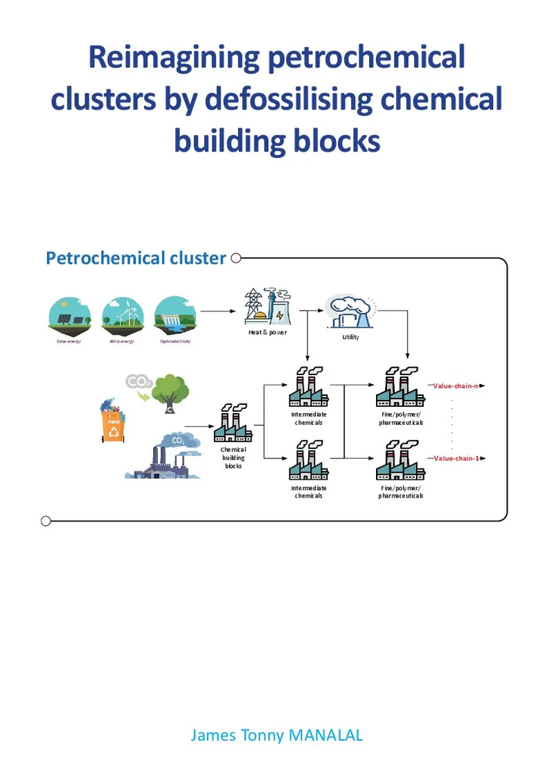
Tonny Manalal
Reimagining petrochemical clusters by defossilising chemical building blocks
18 mei 2026
Overig
Open Ebook
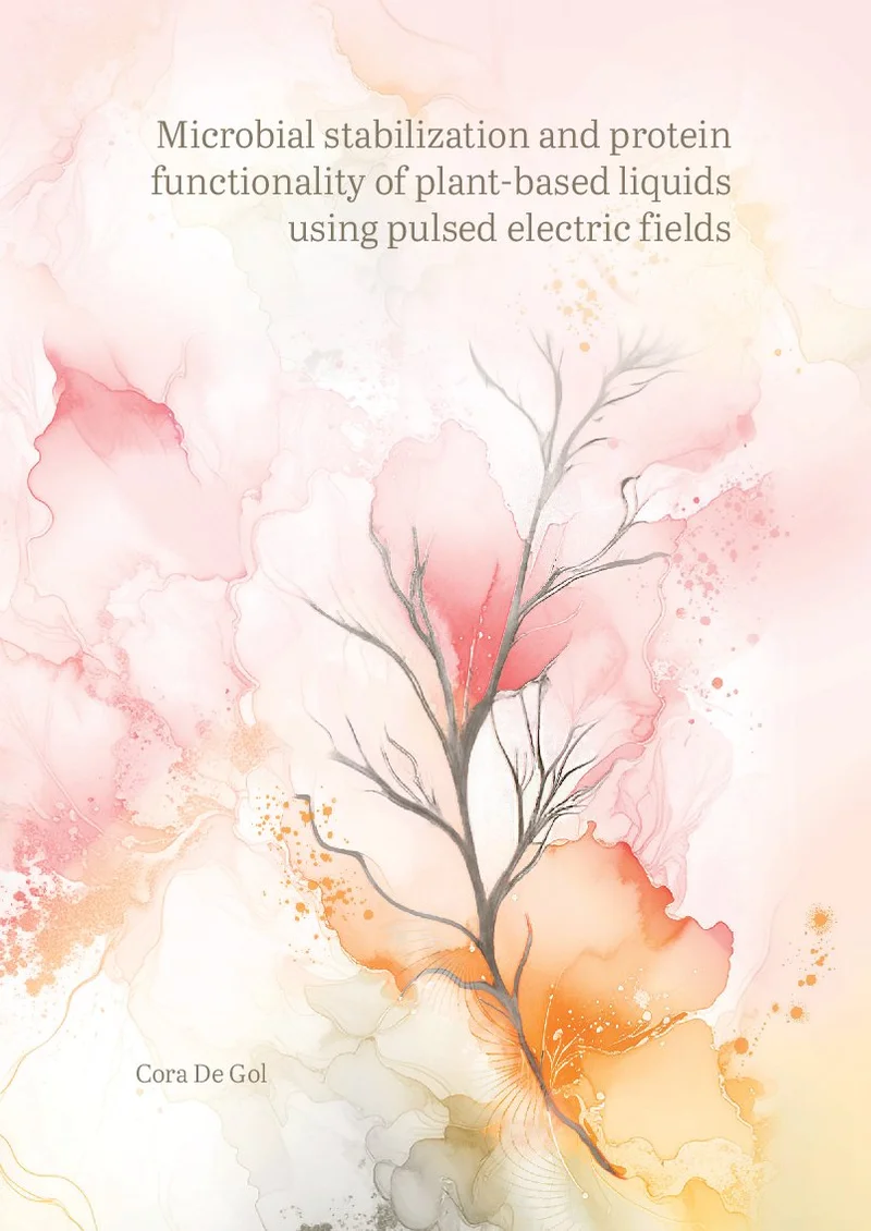
Cora De Gol
Microbial stabilization and protein functionality of plant-based liquids using pulsed electric fields
17 juni 2026
Wageningen University
Open Ebook
We print for the following universities


