Dixit Guleria
Uniaxial Orientation of Polyethylene Films for Recyclable Flexible Packaging
21 mei 2025
TU Eindhoven
Open Ebook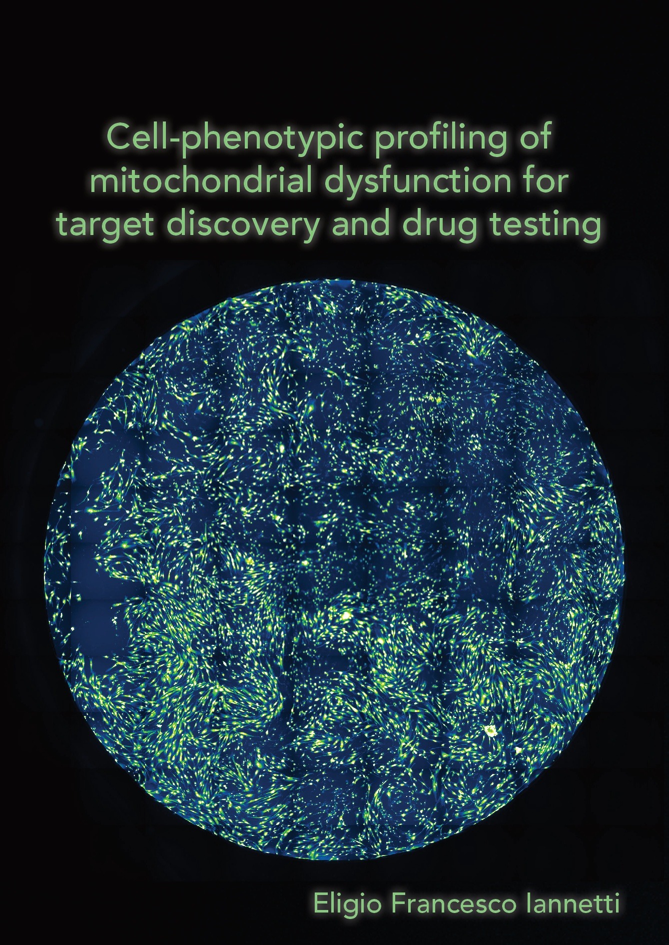
Share this project
Summary
The primary aim of this PhD project was to characterize cellular consequences of mitochondrial dysfunction at the phenotypical level for target discovery and drug testing. In order to do that, I focused on the development of novel automated live-cell imaging technologies and cell-based assays optimization. These were applied mainly to Primary Human Skin Fibroblasts (PHSFs) derived from healthy donors and Leigh Syndrome (LS) patients. LS is a severe neurological disorder usually becoming apparent in the first year of life by progressive loss of mental and movement abilities. This typically results in death within two to three years, usually due to respiratory failure (Gerards et al., 2016). The PHSFs derived from LS patients used in these studies have the most common mitochondrial oxidative phosphorylation (OXPHOS) enzyme defect associated with this disease phenotype, being isolated complex I (CI) deficiency (Fassone and Rahman, 2012; Ma et al., 2013).
Chapter 1 introduced mitochondrial structure, function and dysfunction from the molecular level and up to the clinics including drug development strategies. The importance mitochondria execute in relation to the overall human health status is highlighted. The concept of mitochondrial morphofunction is also introduced discussing the important interaction between structure and function. As morphofunction is a key indicator of the mitochondrial and overall cellular health status, and because of its biological relevance, it was measured or discussed in every chapter of this thesis. The chapter continues introducing the devastating clinical aspects of mitochondrial diseases and in particular CI deficiency and LS that are pathologies with a high unmet medical need. The general introduction ends with an overview on the drugs development process focusing on in vitro preclinical work. In particular cell-based phenotypic screening are presented as the main methodological approach used by pharmaceutical companies for target discovery and drug testing.
To evaluate how mitochondria contributed to disease and to perform preclinical in vitro drug development, a broad collection of technologies exists, both commercially available and in research laboratories. In Chapter 2 we presented a critical review of the literature on how live-cell high content microscopy can be used for image-based phenotypic profiling to assess mitochondrial (dys)function. Comprehensive guidelines about live cell imaging technologies were here presented to introduce the applications developed in the experimental chapters of this thesis. With that perspective, we discussed a selection of live-cell fluorescent reporters and imaging strategies and human cell models and discuss their pros/cons in mitochondrial research. We also presented an overview of live-cell high content microscopy applications used to detect disease-associated cellular phenotypes and perform cell-based drug screening.
Chapter 3 focused more specifically on how mitochondrial morphofunction can be studied combining high-content and high-throughput strategies. In this chapter, different applications which can be used to analyze genetic and/or drug-induced effects at the level of individual organelles, cells and cell populations are briefly discussed. In Chapter 4 we presented a newly developed protocol to perform multiplexed high-content analysis of mitochondrial morphofunction. This allow unbiased and automated quantification of mitochondrial morphofunction, cellular and nuclear parameters in PHSFs. Cells were cultured in 96-well plates and stained with three fluorescent dyes targeting mitochondria, cytosol and nuclei. Multispectral fluorescence images were acquired using automated microscopy and processed to extract 44 descriptors. Subsequently, the descriptor data were subjected to univariate, bivariate and multivariate analysis. Although this protocol was specifically developed for PHSFs, this is transferable to other cell types. In Chapter 5 this protocol was applied to the HT22 neuronal cell model. In this study, the mitochondrial morphofunctional analysis was used to analyze cellular metabolic flexibility using small-conductance Ca2+-activated K+ channels (SK) activation and investigating its phenotypical consequences. HT22 were treated with the chemical compound CyPPA to induce a pharmacological SK activation. In the presence of glucose in the culture medium, many cell models cultured in vitro generate almost all ATP via glycolysis rather than using the mitochondrial OXPHOS system (Crabtree, 1929). Therefore, cell culture medium where glucose is replaced by galactose is a common experimental setting initially developed to induce or enhance pathological mitochondrial phenotypes (Robinson et al., 1992; Rossignol et al., 2004). This strategy enforce ATP production to rely on mitochondrial metabolism, prevent glycolysis upregulation and limit metabolic flexibility (Rossignol et al., 2004). In Chapter 5, in order to limit cellular compensatory mechanisms by the inhibition of glycolytic upregulation, we performed mitochondrial morphofunctional analysis of CyPPA-treated HT22 cells in galactose-based media and compared it with glucose-based media as control condition.
In Chapter 6, in order to induce a metabolic stress, enhancing the PSHF CI deficiency-induced aberrations, we also used the galactose-based medium strategy. We performed a cellular and mitochondrial phenotypical characterization of the mitochondrial diseased cells growing in galactose-based medium and we compared those to the same cells growing in a glucose-based medium. We investigated cell viability, mitochondrial morphofunction, reactive oxygen species (ROS) levels and ATP homeostasis. A panel of six cell lines were used, three derived from healthy donors and three derived from LS patients harboring mutations in nuclear DNA (nDNA)-encoded CI genes (NDUFS7, NDUFS8, NDUFV1). Following optimization of the cell culture protocol and of the glucose-based medium composition, LS fibroblasts died in the galactose medium, whereas control cells did not. LS cell death was dose-dependently inhibited by pyruvate, malate, oxaloacetate, α-ketoglutarate, aspartate, and exogenous NAD+ (eNAD), but not by lactate, succinate, α-ketobutyrate, and uridine. Pyruvate and eNAD increased the cellular NAD+ content in galactose treated LS cells to a different extent and co-incubation studies revealed that pyruvate-induced rescue was not primarily mediated by NAD+. Functionally, in LS cells glucose-by-galactose replacement increased mitochondrial fragmentation and mass, depolarized the mitochondrial membrane potential (∆ψ), increased H2DCFDA-oxidizing ROS levels, increased mitochondrial ATP generation, and reduced the total cellular ATP content. These aberrations were differentially rescued by pyruvate and eNAD, supporting the conclusion that these compounds rescue galactose-induced LS cell death via different mechanisms. These findings establish a cell-based strategy for intervention testing and enhance our understanding of CI deficiency pathophysiology.
Chapter 7, like Chapter 6, also presented a phenotypic and molecular characterization of PSHF CI deficiency-induced aberrations under a stress condition. In this study, redox stress was induced treating the cells with L-buthionine-(S,R)-sulfoximine (BSO) to reduce the concentration of glutathione (GSH), the main cellular antioxidant. A panel of six cell lines were used, three derived from healthy donors and three derived from LS patients harboring mutations in nuclear DNA (nDNA)-encoded CI genes (NDUFS7, NDUFS2, NDUFV1). In respect to Chapter 6, while NDUFS7 cell line was the same one used in both the studies, NDUFS2 was a different cell line mutated in a different gene of CI, NDUFV1 was a different cell line with a different mutation in the same gene of CI. We previously demonstrated that the GSH synthesis inhibitor BSO decreases cellular GSH content to a similar degree in PHSFs from LS patients and control healthy donors. Interestingly, BSO treatment greatly reduced the viability of LS patient cells (all harboring an isolated mitochondrial CI deficiency), whereas healthy controls cells (CT). However, the reason for this differential BSO effect is still unclear. In this last chapter we performed phenotypic and molecular profiling of LS and CT cells to investigate the effect of BSO prior to cell death induction. PHSF incubation with a non-toxic BSO concentration (1 µM) for 24h decreased mitochondrial and cytosolic GSH levels in LS and CT cells in a similar manner. No alterations in mitochondrial morphofunction, cellular ATP content or cellular lipid peroxidation state were observed up to 2 hours prior to cell death induction. However, when lipid peroxidation was measured from 12h to 18h after BSO treatment, an oxidative burst almost concomitant with the loss of cell viability was identified as LS-specific pathological phenotype. We previously demonstrated that KH176m, the metabolite of a new clinical-stage drug candidate KH176, fully inhibited BSO-induced LS cell death. Here we provide preliminary evidence that this cell death is paralleled by increased lipid peroxidation, which is prevented by KH176m. KH176m efficacy was also benchmarked against Ferrostatin-1, the first identified inhibitor of ferroptosis, a lipid peroxidation-dependent cell death. This pilot study highlights a causative role for lipid peroxidation in the BSO-induced specific death of LS patient cells and enhances our understanding of KH176m mode-of-action.