Pawan Kishore Ravindran
Surgical and Radiotherapeutic Nuances in Skull Base Tumors
23 juni 2026
Universiteit Maastricht
Open Ebook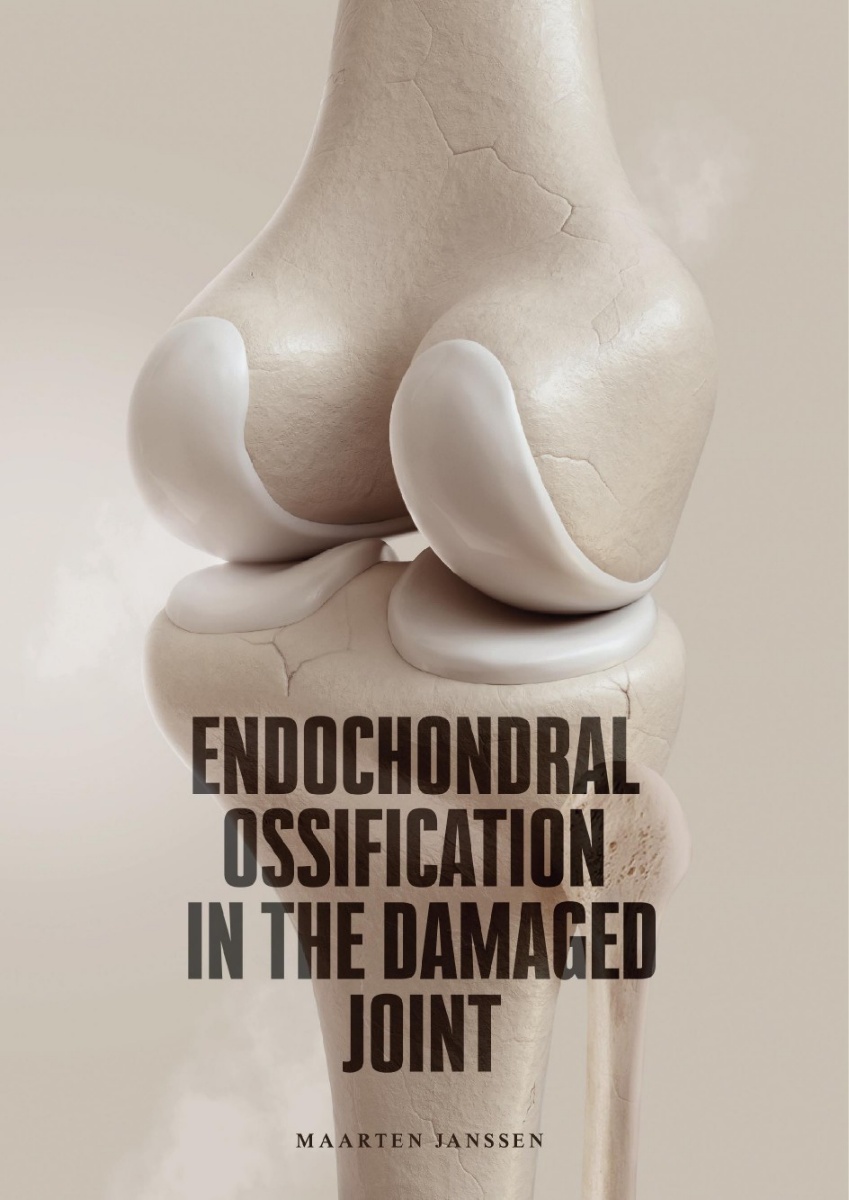
Deel dit project
Samenvatting
De wereldwijde levensverwachting neemt toe en ter verbetering van de kwaliteit van leven wordt lichaamsbeweging geadviseerd, zelfs op gevorderde leeftijd. De positieve effecten van lichaamsbeweging zijn uitgebreid beschreven in wetenschappelijke literatuur. Ironisch genoeg is een mechanisch trauma bij sporten beschreven als de grootste oorzaak van lokale kraakbeendefecten. Kraakbeen heeft weinig regeneratieve capaciteit en een beschadiging van kraakbeen leidt daardoor vaak tot verdere degeneratie. Een belangrijke component in het proces van articulaire kraakbeenschade en degeneratie van het gewricht is de veranderde expressie van biomoleculaire factoren. Deze biomoleculaire factoren beïnvloeden de homeostase van het gewricht en zorgen voor een toename van ongewenste endochondrale ossificatie (EO) van het articulaire kraakbeen en osteofyt vorming. Het werk in dit proefschrift focust op het begrijpen van veranderingen in de rol van EO in focale en diffuse schade aan articulair kraakbeen en op de mogelijkheden om gebruik te maken van de biomoleculaire mechanismen die actief zijn in EO voor de behandeling van beschadigd articulair kraakbeen.
In het eerste deel van dit proefschrift werd de invloed van patiëntkarakteristieken op het resultaat van kraakbeenchirurgie onderzocht, met ook de rol van EO in dit proces. In Hoofdstuk 2 werd een groep patiënten 25 jaar na perichondrium transplantatie (PT) geanalyseerd, waarbij perichondrium van de rib getransplanteerd werd naar de knie. We ontdekten dat het risico op falen van deze ingreep (gedefinieerd als het ondergaan van een grote hersteloperatie) lager was en dat de functie van de knie (gemeten door middel van de International Knee Documentation Committee (IKDC) score) beter was wanneer adequate patiënt selectie werd toegepast. Factoren die het risico op falen significant verhoogden, waren een eerdere operatie aan dezelfde knie en een langere duur van de symptomen voorafgaand aan de kraakbeenoperatie. Een jongere leeftijd ten tijde van de operatie werd geassocieerd met een betere IKDC-score na 25 jaar follow-up. Een morfologische en biochemische evaluatie van het herstelde kraakbeen werd door middel van een 7 tesla (7T) MRI verricht in patiënten van hetzelfde PT cohort en de resultaten werden vergeleken met een cohort patiënten die in dezelfde periode een autologe chondrocyten transplantatie (ACT) hadden ondergaan (Hoofdstuk 3). Er werd geen correlatie gevonden tussen klinische vragenlijsten en de MOCART (Magnetic Resonance Observation of Cartilage Repair Tissue) score of biochemische verslechtering van de kraakbeentransplantaten. In zowel PT als ACT patiënten kwamen intralesionale osteofyten frequent voor. Deze osteofyten kunnen leiden tot biochemisch aantoonbare schade aan het tegenoverliggende tibiale kraakbeen. Deze schade was meer afhankelijk van de morfologie van deze osteofyten (i.e., een osteofyt die tot in het oppervlak van een transplantaat groeit) dan van het percentage verkalking van het transplantaat.
In het tweede deel van dit proefschrift onderzochten we hoe het EO-proces beïnvloed kan worden. In Hoofdstuk 4 toonden we aan dat cyclo-oxygenase-2 (COX-2) inhibitie niet alleen EO in de groeiplaat en in de fractuurcallus verminderde, maar ook de periostale kraakbeenvorming in een in vivo konijnenmodel. De inhibitie van COX-2 werd bereikt door het gebruik van celecoxib, een selectief COX-2 remmend medicijn dat valt in de groep Non-Steroidal Anti-Inflammatory Drugs (NSAIDs). Ondersteund door voorgaande studies van onze onderzoeksgroep die een bifasisch patroon van COX-2 expressie in EO aantoonden, laten deze resultaten zien dat celecoxib niet alleen hypertrofe differentiatie van kraakbeencellen remt, maar ook de chondrogene fase van EO in bepaalde omstandigheden kan remmen. Een andere manier om het EO-proces te beïnvloeden werd onderzocht in Hoofdstuk 5. Het vervaardigen van kraakbeen uit progenitorcellen wordt vaak belemmerd door ongewenste EO. Om het probleem van nadelige hypertrofe differentiatie van chondrocyten tijdens ectopische kraakbeenvorming te voorkomen werd een eerder gebruikte biogel aangevuld met de belangrijke kraakbeeneiwitten aggrecan of cartilage oligomeric matrix protein (COMP). Deze eiwitten bleken in staat te zijn om het proces van ectopische kraakbeenvorming te verbeteren in een in vivo konijnenmodel, door het onderdrukken van hypertrofe differentiatie van het gevormde kraakbeenweefsel.
In het laatste deel van dit proefschrift werden potentiele behandelopties onderzocht die het intra-articulaire milieu kunnen verbeteren om gewrichtspijn te verminderen en progressie van kraakbeenschade naar artrose uit te stellen of te voorkomen. De huidige beschikbare orale medicamenteuze behandelingen hebben vele nadelen die kunnen worden overwonnen door intra-articulaire toediening van geschikte medicatie. Het medicijn blijft echter vaak maar een korte tijd in het gewricht aanwezig en er is een medicijnafgiftesysteem nodig om de effectiviteit van intra-articulaire therapie voor kraakbeenschade te verbeteren. Verschillende soorten medicijnafgiftesystemen zijn: Verlengde afgifte systemen, gecontroleerde afgifte systemen of autoregulatoire systemen. De literatuurstudie verricht in Hoofdstuk 6 liet zien dat medicijn-afgiftesystemen gemaakt kunnen worden van verschillende materialen. Sinds het gebruik van polymeren voor de ontwikkeling van medicijnafgiftesystemen is er veel progressie geboekt. Het gebruik van medicijnafgiftesystemen voor de behandeling van artrose is veelbelovend, maar er is nog geen klinische trial die aangetoond heeft dat deze medicijnafgiftesystemen in combinatie met medicijnen in staat zijn het ziekteproces van artrose te veranderen. Hoofdstuk 7 van dit proefschrift beschrijft de ontwikkeling en het testen van de polyester amide-celecoxib (PEA-CXB) micropartikel als medicijnafgiftesysteem. De farmacokinetische eigenschappen en reactie op een inflammatoire (artrotische) omgeving werden in vitro onderzocht in cellysaten verkregen van een neutrofiele HL-60 cellijn. Vervolgens werden de bio-compatibiliteit en degradatie van de PEA-CXB micropartikel getest in een in vivo rat model waarbij post-traumatische artrose was geïnduceerd door middel van het doornemen van de voorste kruisband en gedeeltelijke verwijdering van de mediale meniscus. De PEA-CXB micropartikels veroorzaakten geen detecteerbare bijwerkingen en waren geschikt als medicijnafgiftesysteem. Een verandering van het artroseproces door de PEA-CXB micropartikels werd niet aangetoond. In rattenknieën met artrose was er een toegenomen degradatie van de micropartikels en de toevoeging van celecoxib aan de partikels zorgde voor een afname van dezelfde degradatie, wat toebedeeld werd aan auto-regulatoire eigenschappen van het medicijnafgiftesysteem. In Hoofdstuk 8 werden de belangrijkste bevindingen van dit proefschrift in breder perspectief geplaatst.
De wereldwijde ziektelast van artrose stijgt snel, met name in relatief jonge patiënten. Wanneer een totale knieprothese wordt geplaatst bij jonge patiënten is het risico op een revisie operatie groot. Daarom is er meer aandacht nodig voor gewrichtssparende behandelingen. Voor een succesvolle gewrichtssparende behandeling zijn een tijdige herkenning, patiëntselectie en behandeling van kraakbeenschade en artrose nodig. Een adequate behandeling kan bestaan uit leefstijl, farmacologische en/of chirurgische interventies. De bevindingen in dit proefschrift tonen dat de verbetering van gewrichtssparende behandelingen bereikt kunnen worden op verschillende niveaus en vanuit verschillende perspectieven (bijvoorbeeld vanuit de cel en vanuit de patiënt). Echter, het belangrijkste voor een succesvolle gewrichtssparende behandeling van patiënten met gewrichtsschade is een verandering in de benadering van het klinische probleem door orthopedisch chirurgen, andere zorgverleners en wetenschappers. Een benadering waarbij de “patient journey” van een persoon met een gezond gewricht tot aan een gewrichtsvervanging gevolgd en begeleid kan worden door middel van de juiste interdisciplinaire samenwerking. Hierdoor wordt de juiste zorg op de juiste tijd en plaats geboden om mensen en patiënten in beweging te houden en zo andere chronische ziekten te voorkomen.
Impact paragraph
Cartilage is a durable, but flexible tissue that occurs throughout the body. In articular joints, hyaline cartilage comprises a layer that covers the ends of the bones and provides a surface with very low friction that makes movement possible and at the same time functions as a shock absorber. Unfortunately, cartilage has a very low healing capacity. Therefore, damage to articular cartilage is not resolved and often leads to a pathway of joint deterioration and finally osteoarthritis (OA). Articular cartilage lesions are found in up to 62% of the knees of adults without any symptoms of joint pathology. When cartilage degradation becomes symptomatic, or even progresses into OA, this can have an enormous impact on a person’s life. Osteoarthritis is a leading cause of disability worldwide and its burden is only expected to increase due to the ageing population and increasing incidence of obesity. Furthermore, disabling OA leads to a substantially reduced long-term work participation and is therefore a major economic concern as well. The most frequently applied therapy for end-stage OA is arthroplasty, but the results of total knee arthroplasty (TKA) in working patients are dissatisfying and one third of patients does not return to work after TKA. The lifetime risk of implant revision is increased in younger patients (up to 35% for men in their early 50s). In addition, the median time to revision is significantly shorter in patients who were younger than 60 at the time of TKA. It is thus of great social and economic value to prevent, or at least postpone progression towards end-stage OA and subsequent (early) TKA. Therefore, the main goal of this thesis was to elucidate how the process of endochondral ossification (EO) can be influenced to improve the treatment of damaged cartilage (i.e., focal cartilage defects and OA).
Conclusion of main findings
The process of EO is an essential factor in cartilage damage and repair. The findings in this thesis confirm that patient characteristics can negatively influence the outcome of cartilage repair surgery. Potentially by impairing the joint homeostasis and increasing joint inflammation and subsequent EO of the repaired cartilage tissue. The work in this thesis underlines the potential of inhibiting inflammation and influencing the EO pathway with the aim to improve cartilage repair and OA treatment by reducing undesired chondrocyte hypertrophy.
Implications for research
The influence of inflammation and patient characteristics on the outcome of cartilage repair surgery and OA treatment is widely recognized, but still not fully understood. The data in this thesis demonstrate that adequate patient selection can improve the outcome of cartilage repair surgery. In addition, the added value of 7T MRI is underlined. The detailed visualization of morphological and biochemical differences (such as increased calcification of repaired cartilage) suggests that inflammation and EO can influence the results of articular cartilage repair. This shows that innovations in imaging, such as (high-field) MRI, can aid in an increased understanding of the mechanisms of treatment failure and subsequently provide directions to improve treatment strategies. However, the influence of EO on the quality of cartilage repair tissue and subsequent clinical outcome should be further elucidated in prospectively designed studies. Increased knowledge on the use of 7T MRI also provides a way to evaluate articular cartilage non-invasively and at multiple timepoints, facilitating future clinical research on the influence of EO on articular cartilage damage and repair. This future research could elaborate on the work presented in this thesis that describes the potential improvement of ectopic cartilage tissue formation by influencing inflammation and EO via the inhibition of cyclooxygenase (COX)-2. In addition, the chondrocyte hypertrophy-suppressive effect of aggrecan and cartilage oligomeric protein (COMP) without impairing cartilage formation provides an interesting starting point for future studies.
Implications for individual patients and society
Good surgeons know how to operate, better ones when to operate, and the best when not to operate. This was stated in a BMJ editorial dating back to 1999, but is still applicable. Not performing unnecessary surgical procedures protects patients from avoidable strain. In addition, it decreases hospital costs and all other socioeconomic costs involved with the surgery. Key findings in this thesis increased the knowledge on risk factors and adequate diagnostic tools to detect cartilage defects and (early) OA. Early detection of cartilage damage provides the opportunity to improve the ‘patient journey’ by starting early with a suitable treatment, preserve a functional joint and prevent loss of mobility in patients. This can subsequently avoid costly procedures in progressed OA such as revision of total knee arthroplasty or socioeconomic costs caused by disability in patients of working age. Furthermore, an increased understanding was obtained on the role of inflammation and EO on the treatment of cartilage damage and OA by the development of the PEA-CXB microsphere. Derived strategies could further elaborate on the inhibition of chondrocyte hypertrophy and inflammation to treat cartilage damage and potentially lead to a reduction of the amount of (early) TKA and subsequent revision TKA. Next to reduced socioeconomic costs, a reduction in the amount of (early) TKA can also decrease secondary (psychological) complaints and improve the quality of life of OA patients.
Implications for health care professionals
Next to the implications for the individual patient and society described above, the research results presented in this thesis are valuable for health care professionals as well. We found that late and multiple surgeries in older patients decrease the chance of success in focal cartilage repair surgery. This underlines the importance of adequate early treatment of articular cartilage damage to prevent further deterioration of the joint. This can be achieved by educating primary physicians to recognize patients with possible articular cartilage damage that are suitable for early referral to an orthopaedic surgeon. This might be even more applicable for physical therapists, as in the Dutch health care system, physical therapy is often the first line of treatment for patients with (minor) musculoskeletal complaints. Orthopaedic surgeons can benefit from a timely referral and potentially provide less invasive, joint preserving treatments.
Communication towards health care professionals
A timely treatment of articular cartilage damage can prevent further deterioration of the joint. In addition, the findings in this thesis showed that a timely treatment of cartilage damage decreases the risk of treatment failure and subsequent TKA. To facilitate this timely treatment of articular cartilage damage, primary healthcare providers involved in the treatment of patients with focal cartilage defects or (early) OA have to be taught that early referral can be joint-preserving. To educate health care providers, publishing research results in peer reviewed journals is essential, but is not enough. Medical information is abundantly available on the internet. However, keeping an overview is complex and the abundant information is impossible to interpret and apply for all different health care providers. For the results of this thesis (and other research) to consistently reach all relevant health care providers, communication will have to be improved. In the following years there will have to be significant advancements in the infrastructure of electronic health records. The currently, not directly linked electronic health records of (amongst others) primary physicians, physical therapists and orthopaedic surgeons will have to be linked or integrated so that all health care providers can have the access to relevant information and are provided with adequate feedback on their treatment actions, ideally supported by scientific research. This can be facilitated by the use of a personal health environment in which personal medical data is owned by the patient and can be shared with different institutions.