Zjala Ebadi
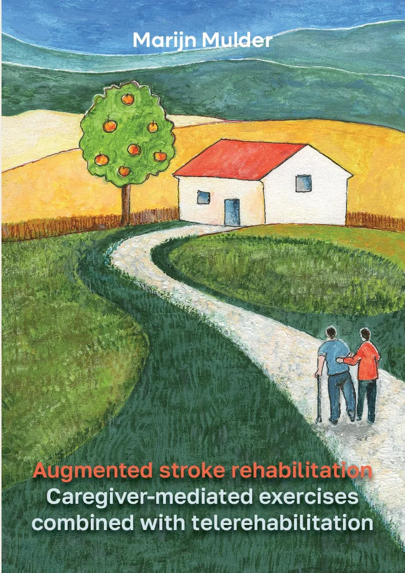
Persistent Fatigue in Chronic Respiratory Conditions
14 september 2026
Radboud Universiteit
Open Ebook
Share this project
Summary
Summary and Future Perspectives
In recent years, biomedical imaging research has profoundly been influenced by the advent of automated image analysis methods, currently primarily based on machine learning methodologies. At the same time, innovations in cerebral endovascular interventions have revolutionized stroke treatment practices. These two developments have led to a unique opportunity to further optimize treatment and personalized care in acute ischemic stroke. This thesis combined and advanced the latest technical and clinical research in this domain, by focusing on automated and quantitative analysis of peri-operative cerebral angiography for diagnosis, interventional decision-support, and prediction of functional outcome.
Parts I to III address several technical challenges in the image analysis of digital subtraction angiography, ranging from image acquisition and image processing to image interpretation. Correspondingly, Part IV presents further clinical validations in connection to these technical contributions.
During endovascular procedures in stroke, multiple 2D DSA time series are collected. The quality of cerebral DSA is often negatively impacted by head motion during acquisition, leading to decreased diagnostic value. In Chapter 2 we presented AngioMoCo, a deep learning-based strategy towards motion-free digital subtraction angiography. Traditional approaches to motion correction rely heavily on non-rigid registration techniques and employ sparse key points and non-rigidity penalties to mitigate vessel distortion. However, these methods are time-consuming. Recent learning-based methods alleviate subtraction artifacts by predicting the subtracted frame from the corresponding unsubtracted frame, but do not explicitly compensate for motion-induced misalignment between frames. This impedes the serial evaluation of blood flow, and often causes unwanted alterations in the appearance of vasculature and contrast flow dynamics. AngioMoCo leverages a contrast extraction module to disentangle contrast flow from body motion and a deformable registration module to concentrate on motion-induced deformations. In such a way, the registration focuses on correcting patient motion rather than erasing intensity changes caused by contrast flow. This strategy improves registration quality while being orders of magnitude faster than iterative elastix-based methods. We demonstrated on a large national multi-center dataset (MR CLEAN Registry) that AngioMoCo achieves high registration accuracy while preserving vascular features, improving the quality and clinical utility of DSA for diagnosis and treatment planning in endovascular procedures.
Apart from misalignment between frames within a DSA series, misalignments often occur between DSA series acquired at different time points of a single patient. This limits the direct comparison of treatment effects on cerebral vasculature. Chapter 3 investigated approaches to automatically align cerebral DSA series. Transformations that account for differences in scale prove effective in aligning cerebral DSA series. By precisely identifying landmarks through deep learning techniques, registration of pre-/post-EVT DSA series can be performed using traditional point-based methods, achieving a success rate of 85%. This level of success holds across patients with various occlusion locations and procedural outcomes. These findings may enable further automation of DSA image analysis and procedure evaluation, contributing to outcome prediction and procedural decision-making for EVT.
Vessel segmentation is another heavily researched topic in medical images of anatomies such as eyes, brain, lungs, and heart. In the context of cerebral DSA, it is clinically relevant to extract and distinguish arterial and venous vessels for potential biomarker extraction. Part II of the thesis described vessel segmentation methods tailored to 2D DSA time series. Chapter 4 used the Frangi filter and thresholding to perform vessel segmentation and separated arteries and veins using unsupervised machine-learning methods based on features derived from the time-intensity curves. As a further improvement, Chapter 5 proposed a fully automatic deep learning-based artery-vein segmentation method. It integrates both spatial vascular appearance and temporal contrast flow dynamics in a unified end-to-end framework, producing high-quality multi-class segmentations from cerebral DSA series with variable lengths. The method promises to facilitate vessel-based quantitative analyses for clinical diagnosis, outcome prediction, and treatment planning in endovascular interventions.
While the previous chapters focus on general image processing techniques in DSA, Part III delves into specific clinical applications using quantitative image analysis methods. Chapter 6 presented an explainable, robust, and fully automatic perfusion quantification method, autoTICI. This was to address the limitation of TICI and other existing treatment evaluation metrics in acute ischemic stroke. Existing TICI scores are defined in coarse ordinal grades based on visual inspection, leading to inter- and intra-observer variation. In contrast, autoTICI is an automatic, reproducible, and continuous reperfusion percentage. On the MR CLEAN registry data, autoTICI demonstrated good correlations with the extended TICI (eTICI) scored by experienced neuroradiologists and showed comparable capability in predicting patient functional outcomes at 3-month following the EVT procedure, revealing its potential in future studies and clinical practice.
During an EVT, the interventionalist retracts the thrombus whereby navigating to the clot location, advancing through the thrombus, and retrieving the thrombus can involve changing guide wires several times. Adverse events could occur during the procedure, which may necessitate immediate therapeutic responses. Chapter 7 presented the first study that addresses automatic intracranial vessel perforation detection during EVT through the use of spatio-temporal networks. Employing data collected from multiple (inter-)national databases, the developed deep learning models demonstrated expert-level performance on par with that of expert human evaluators and good generalization capability, revealing its potential value in assisting therapeutic decision-making in clinical practice.
While Chapters 6 and 7 provides concrete examples of how quantitative evaluation could help in image-guided EVT, more quantitative biomarkers could be measured and compared across patients if such parameters are independent of the contrast injection profile during the acquisition of DSA. In a swine study, Chapter 8 investigated the quantitative properties of deconvolution-based perfusion angiography. It confirms the superior independence and sensitivity of deconvolution-based perfusion angiography (CBV DSA, CBF DSA, Tmax, MTT DSA) over parameters that are directly derived from the time-intensity curves (AUC, peak intensity, TTP) in DSA under varying injection protocols between acquisitions. These findings may further facilitate quantitative assessments of perfusion angiography in endovascular interventions.
Finally, Part IV described extensive clinical investigations regarding the proposed methods and adverse events during EVT procedures. Chapter 9 studied the performance of autoTICI in a larger sample size, including 467 patients in the MR CLEAN Registry with an ICA, M1 and M2 occlusion. AutoTICI proves to have similar predictable applications as eTICI when adjusting for prognostic factors and could be used as an alternative to visual reperfusion assessment to improve reproducible and uniform reporting when imaging requirements are met.
Although challenges remain to be addressed before automated learning methods become a standard feature in clinical practice, it is evident that these techniques hold substantial potential to make a meaningful impact on the diagnosis and treatment of acute ischemic stroke.
Future perspectives
The stroke management process involves pre-treatment diagnosis, acute treatment, and rehabilitation. The objectives outlined in the introduction of this thesis, described from the perspective of clinicians and patients, primarily focus on improving acute treatment based on AI and quantitative image analysis techniques. They translate into A) the evaluation of treatment success (Goal 1, 3, and 4 in Section 1.7), B) therapeutic decision support (Goal 1 and 2), and C) extraction of quantitative biomarkers to predict patient recovery (Goal 3 and 4). This thesis has touched upon all these objectives in various aspects. In terms of treatment evaluation, Chapter 6 introduced autoTICI, which is the first automated and interpretable brain reperfusion scoring framework. This framework offers an objective and granular approach to quantify brain perfusion. Subsequently, Chapter 9 validated the proposed AI-based method using large clinical data derived from multi-center clinical studies and comprehensively discussed its potential and limitations for clinical translation. Concerning therapeutic decision support, Chapter 7 delved into the challenge of detecting interventional complications, i.e., cerebral vessel perforations. The proposed method demonstrated robust detection within 2D time series by harnessing the temporal characteristics of cerebral contrast flow in spatio-temporal learning. Regarding quantitative biomarkers, this thesis presented preclinical in-vivo swine studies to explore the quantitative properties of DSA in brain perfusion analysis. The findings reveal future directions for automated quantitative analysis in DSA, contributing to the identification of quantitative clinical biomarkers and offering prospects for a wide range of clinical applications. Besides, the technical developments on registration (Chapters 2 and 3) and segmentation (Chapters 4 and 5) could also assist in quantitative biomarker extraction in DSA.
This thesis addresses several technical and clinical challenges in image-guided EVT for stroke while also revealing ample opportunities for further exploration of those topics. In terms of treatment evaluation, many existing deep learning methods often confine their use cases to proximal occlusion locations, such as the ICA and/or M1 segment ([47, 147, 148]). To enhance clinical relevance, future studies may further generalize such methods in diverse clinical scenarios, and leverage clinical knowledge priors to provide a comprehensive assessment of the functional independence of patients. With the infusion of AI, it may even be valuable to explore novel quantitative metrics beyond TICI for assessing treatment success that consider the functional status of affected brain regions. Regarding therapeutic decision support, we addressed timely detection of procedural complications, but there is a further need for improved peri-procedural visual feedback. Examples include integration of pre-operative images during interventions and registration of images acquired at different time points to visualize treatment effects. These applications promise more efficient treatment timelines and enhanced success rates. For quantitative biomarkers, there is also great potential for further exploration. Despite significant technical advancements in recent years, about half of stroke patients who undergo successful reperfusion treatments still struggle to regain long-term functional independence in their daily lives. We do not fully understand why this happens yet. To better understand this phenomenon and develop improved treatment options, we need to further explore quantitative imaging features, study their relevance for improved therapeutic decision-making and outcome prediction, and their translation into functional indicators in patient recovery.
Beyond the topics presented in this thesis, there are numerous challenges that AI may address in the stroke management process. While this thesis primarily centers on the image analysis of cerebral angiography for EVT, this treatment is not the only stage in the stroke management process. Before treatment, AI-based multimodal image analysis could aid with personalized treatment selection. Beyond evaluating treatment effectiveness, the research community could further improve long-term functional outcome prediction, enabling early intervention and enhancing the overall quality of life for stroke patients. Additionally, future research may explore AI-assisted personalized rehabilitation planning and addressing the expectations of both patients and their families.
The primary objective of our technological innovations and clinical validations is to expedite the evolution toward an enhanced AI-assisted stroke management pipeline. However, there exist several challenges to be addressed for the integration of AI-based image analysis solutions into clinical practice. Below I present a few technical aspects of these challenges, including the need for standardized data acquisition and preprocessing, fostering open science practices, and ensuring the transparency and interpretability of deep learning models.
In the age of deep learning, data is key. However, we need to acknowledge the substantial heterogeneity in data selection and preprocessing procedures, concerning image quality, annotations, acquisition methods, and storage formats. A considerable portion of the dataset is often excluded due to various factors, including the absence of DSA acquisitions, data corruption, incompleteness, suboptimal image quality, and images failing to depict the full areas of interest. These challenges introduce complexity to the development of resilient deep learning algorithms. In my view, we can confront this general challenge by either 1) standardizing the workflow for data acquisition and preprocessing, or 2) developing algorithms capable of handling and learning from heterogeneous, unstructured data. A promising avenue toward standardized image utilization and accelerated algorithm development is through the adoption of open science practices. By sharing knowledge and resources, we can eliminate the need for redundant efforts and work collaboratively toward continuous enhancements. Aligned with this purpose, we are working on initiating an open toolbox for deep learning in cerebral angiography and beyond, where researchers could share, reuse, contribute, and establish standardized practices for image acquisition and utilization.
While data serves as the lifeblood of AI, the issue at hand is typically not data shortage, but rather the underutilization of the data at our disposal. Within the stroke treatment process, a wealth of data is collected, including patient medical histories, clinical metrics, multiple imaging modalities for diagnostic and interventional purposes, and data collected at different time points or patient conditions. Unfortunately, we often do not make the most of this information. For example, pre-operative 3D images (e.g., CTA scans) are predominantly employed for diagnostic purposes, with their value underutilized during the course of EVT. In parallel, peri-operative DSA images often fall short in providing comprehensive insights due to their 2D projective nature. As another example, the challenge of DSA image registration arises, as the 3D rigid motion of the head is not preserved in the 2D projective DSA format. Leveraging the continuously available 3D pre-operative CTA scans, DSA image registration may be enhanced by utilizing 3D CTA as an intermediary agent. The integration of multi-modality data may improve disease modeling, while multi-temporal data uncovers temporal changes in patient conditions. Ultimately, cross-modality learning and data fusion will become increasingly practical. There is a lot more to gain by making the most of the diverse data available for stroke treatment.
The explainability of AI solutions becomes vital as we aim to translate them into clinical practice. As AI progresses into real-world applications, novel deep learning-based medical applications are being increasingly developed in the context of the stroke management process. Nevertheless, only a few have made a successful path towards FDA AI-approved medical software. One of the key concerns is explainability ([233]). In Chapter 6, we proposed a step-by-step deep learning-based framework, as opposed to end-to-end strategies, to quantify brain reperfusion after endovascular therapy in stroke patients. With recent advances in explainable AI, it may be worth exploring transparent end-to-end learning strategies such as conditional generative models ([234, 235, 236, 237]), graph neural networks ([238, 239]), or other methods with enhanced human-interpretibility ([240, 241]).
Current AI techniques excel at extracting valuable insights from extensive datasets. In contrast, clinical experts possess unparalleled skills in continuous learning and logical reasoning. In the foreseeable future, both AI and domain knowledge will coexist. As healthcare expenses rise, data volumes expand, and the time available to clinicians for each patient reduces, AI-based solutions will prove to be a cost-effective complement. They efficiently digest the overwhelming amount of data and furnish essential insights that clinical experts rely on. This may foster a collaborative partnership between clinicians and AI solutions, enabling them to leverage their unique strengths.
To conclude, this thesis presented innovative image analysis methods in cerebral angiography for stroke interventions and subsequent clinical investigations and validations. The presented methods and findings hold promise in the AI-assisted stroke management pipeline. As we look forward, embracing data standardization, open science, and explainable AI techniques would expedite the clinical integration of AI solutions.