Share this project
Andrea Brum
Erasmus Universiteit Rotterdam
Publication date: 20 maart 2020
University: Erasmus Universiteit Rotterdam
ISBN: 978-94-6380-735-7
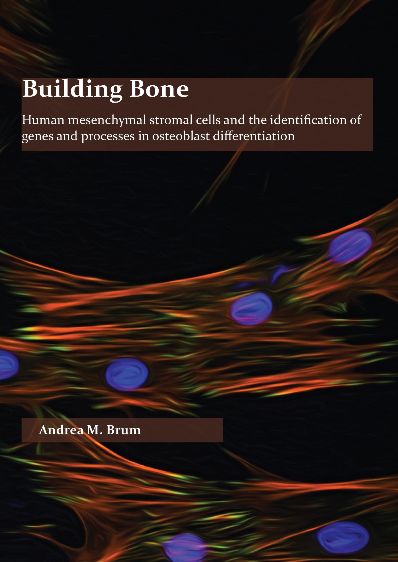
Building Bone
Summary
Bone is a dynamic organ that throughout life undergoes constant remodeling controlled in a balancing act between removal of old bone by osteoclasts and formation of new bone controlled by osteoblasts, as well as its terminally differentiated form the osteocyte. The process of building bone starts with the mesenchymal stromal cell (MSC), a multipotent cell that has the ability to differentiate into a number of cell types including osteoblasts, chondrocytes, and adipocytes. A number of key factors and signaling pathways controlling osteoblast differentiation and activity have been identified over the last few decades; however, a better understanding of the signaling network, their intricate interactions, and what other genes and pathways are involved in osteoblast function is required to develop approaches to enhance bone formation and promote fracture healing for patients with disorders such as osteoporosis. The overall aim of this thesis, through a combined use of bioinformatic, genomic, molecular, and proteomic approaches, was to identify novel factors (compounds, genes, and processes) involved osteoblast differentiation and bone formation to expand the basic knowledge of osteoblast differentiation which can ultimately be used the development of a novel bone anabolic treatment for conditions such as osteoporosis.
In chapters 2 and 3 we demonstrated the use of the web-based Connectivity Map (CMap) tool to find compounds with correlating gene expression profiles to that of human MSCs (hMSCs) undergoing osteoblast differentiation that can affect osteoblast differentiation. The CMap identified parbendazole as the top positively correlating compound and in chapter 2 we showed that parbendazole, independent of an additional osteogenic stimulus, was able to stimulate in vitro human osteoblast differentiation as evidenced by increased ALP activity, mineralization and upregulation of genes important in osteoblast differentiation and extracellular matrix production. Mechanistically, it was found that the osteogenic effect of parbendazole occurs independent of glucocorticoid receptor signaling, but rather via affecting microtubule formation, cytoskeletal organization, focal adhesion distribution, and BMP2 activity. In chapter 3, three additional positively correlated compounds, withaferin A, calcium folinate, and amylocaine, which stimulate osteoblast differentiation and mineralization of hMSCs in vitro were identified. Conversely, compounds with negatively correlated gene signatures to that of differentiating osteoblasts, the results of which we hypothesized may reveal interesting new genes and processes in the osteoblast differentiation process, were examined. Three compounds, salbutamol, metaraminol, and diprophylline, which exhibit a gene signature negatively correlated to our osteogenic gene signature were identified, but only one of these drugs, salbutamol, inhibited dexamethasone-induced osteogenic differentiation of hMSCs were identified Finally, the differentially expressed genes behind the CMap identified compounds and used this approach to find and validate two genes, HMOX1 and STC1, as important factors for human osteogenesis were analyzed.
In chapter 4, CLIC3 was identified as a new gene specifically regulated in the osteogenic lineage of differentiating hMSCs. Lentiviral transduction-mediated overexpression and silencing of CLIC3 during osteogenesis revealed a crucial function for CLIC3 in promoting osteoblast mineralization. Overexpression of CLIC3 in hMSCs strongly enhanced in vivo bone formation in a mouse model for ectopic human bone formation further emphasizing that CLIC3 plays an important role in human osteoblast differentiation. Bioinformatics analysis of proteins identified by CLIC3-His pull down suggests CLIC3s role during osteoblast differentiation may be related cytoskeletal associations and signaling, cell adhesion, and/or nuclear pore formation or transport through. Finally, it was identified that CLIC3 interacts with NEK9 and PTDSS1 during osteoblast differentiation, and inhibition of the NEK9 and PTDSS1 expression reduces osteogenic differentiation of hMSCs.
Chapter 5 describes a novel and temporally shifting role for Muc1 in bone biology. It was showed that deletion of Muc1 in female mice leads to decreased trabecular bone volume in 8-week-old compared to wild type (WT) females; however, this difference disappears by 16 weeks. At the same time, endocortical bone formation rate and femoral stiffness are increased at 16 weeks of age in Muc1 deficient female mice, with a higher rate of endocortical bone formation persisting to 52 weeks of age in Muc1-/- mice. Histomorphometric analysis demonstrated that femurs of 16-week-old Muc1-/- female mice displayed lower numbers of osteoblasts lining the bone surface, while femurs from 8- and 52-week old KO and WT mice did not differ in their number of osteoblasts.
In conclusion, this thesis has presented a number of novel findings in the bone biology field and highlights the complexity of the biology and study of osteoblast differentiation. These studies, and studies like it, are vital in gaining greater knowledge about MSC lineage decision making and osteoblasts differentiation, which is required to develop much needed bone anabolic therapeutics. The findings presented in this thesis highlight the importance of the cytoskeleton in regulating and influencing osteoblast differentiation, and may hold promise as novel anti-osteoporotic treatments, with further research. The discovery of a number of novel factors affecting osteoblast differentiation, including the CMap identified compounds and CLIC3 and MUC1, opens up new avenues in the bone biology field for development of bone anabolic therapeutics.
See also these dissertations
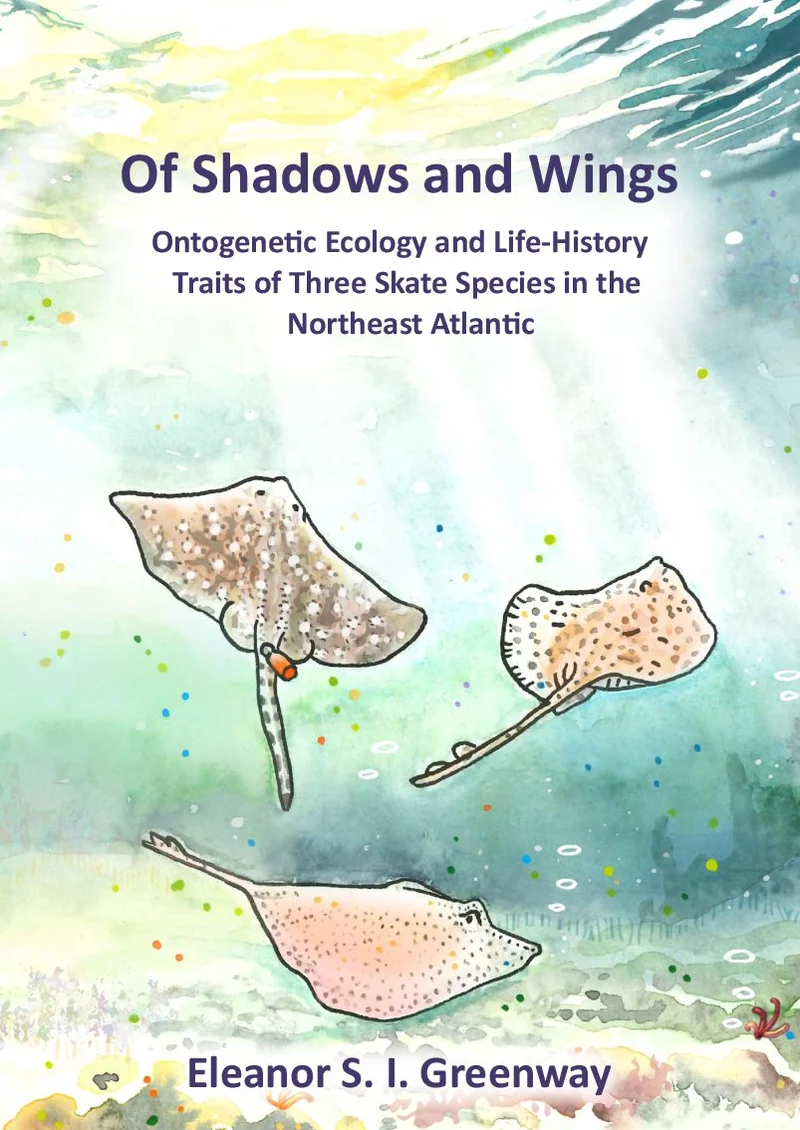


Karlijn Doorenspleet
Improving North Sea biodiversity monitoring using novel molecular approaches
25 juni 2026
Wageningen University
Open Ebook
Haojie Lu
Omics Studies of Cardiometabolic and Skeletal Traits
25 juni 2026
Erasmus Universiteit Rotterdam
Open Ebook
Evans Mudibo
Interaction between acute illness and malnutrition in children in sub-Saharan Africa and South Asia
24 juni 2024
Wageningen University
Open Ebook
Anniek Stuut
The Balancing Act of Allogeneic Haematopoietic Stem Cell Transplantation
10 juli 2026
Universiteit Utrecht
Open Ebook
Danny van den Eertwegh
Charge Transport and Bubble Dynamics in Electrolysis Applications
17 juni 2026
TU Eindhoven
Open Ebook
We print for the following universities


