
Pawan Kishore Ravindran
Surgical and Radiotherapeutic Nuances in Skull Base Tumors
23 juni 2026
Universiteit Maastricht
Open Ebook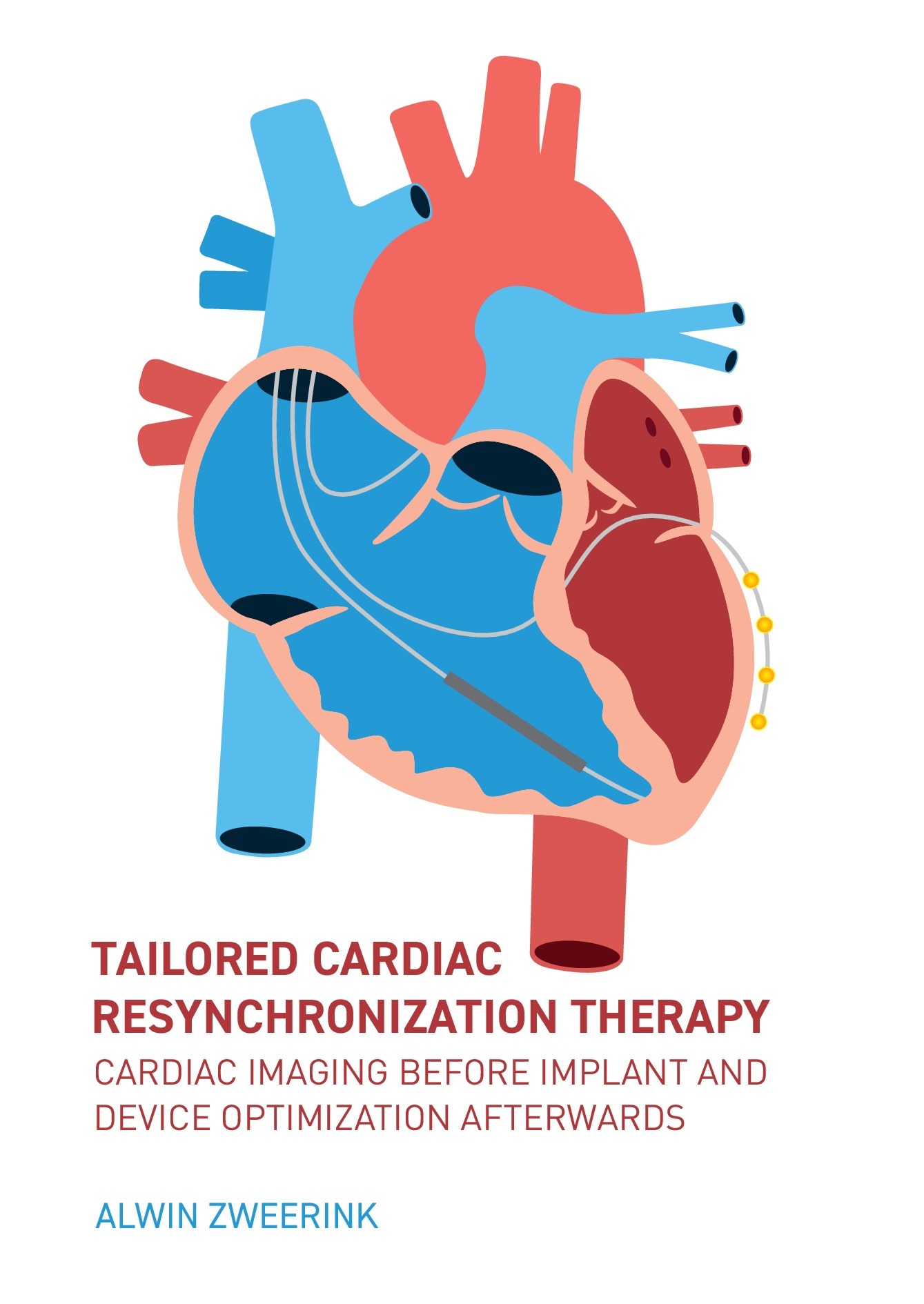
Share this project
Summary
Detection of mechanical discoordination (systolic stretching of segments) rather than dyssynchrony (regional timing differences between segments) yields additional value in the prediction of LV pump function improvement after CRT remains to be proven. Part 1B of this thesis evaluates the role of myocardial strain imaging to improve patient selection for CRT. In chapter four regional strain measurements are combined with LV pressure curves to calculate myocardial work distribution.(4) Contribution of the septum to total LV work varies widely in CRT candidates with LBBB, and the lower the septal contribution to total myocardial work (or the higher the septal waste) at baseline the higher the acute improvement in pump function that can be achieved during CRT. These results are confirmed in a subsequent study by Vecera et al. who showed that wasted septal work strongly predicted CRT response after one year.(5) Myocardial strain imaging therefore provides an insight in the negative effect of LBBB on myocardial work and energy utilization, and reflects the potential benefit that can be achieved by CRT.
In our small-scale study, all patients underwent CMR tagging (CMR-TAG) to obtain high-quality regional strain curves. Availability of CMR-TAG, however, is very limited in clinical practice. Therefore, the role of other non-invasive imaging techniques such as CMR feature tracking (CMR-FT) and speckle tracking echocardiography (STE) is of interest. Chapter five provides a comparison of strain values obtained with CMR-FT and STE versus ‘gold standard’ CMR-TAG. Twenty-seven CRT-candidates, prospectively included in the Markers And Response to CRT (MARC) study, underwent CMR imaging and echocardiographic examination. Both CMR-FT and STE techniques showed to be potentially valuable alternatives for CMR-TAG, especially in the evaluation of mechanical discoordination.
Subsequently, chapter six provides echocardiographic follow-up data in these patients allowing to compare predictive value for CRT response of different strain parameters using multiple imaging techniques. Of all strain parameters, measuring end-systolic septal strain (ESS sep) showed strongest relation with CRT response after one year, irrespective of imaging technique. ESS sep reflects fiber length change of the septum throughout systole. Detection of septal discoordination with higher ESS sep values (i.e. septal stretching instead of contraction) at baseline was associated with more extensive reverse remodeling after CRT. Moreover, measuring ESS sep by any available imaging technique showed to be additive to present guideline criteria (QRS morphology; QRSd). The application of strain imaging has yet not been included in daily practice, but it is likely to become a useful application when evaluating heart failure patients for CRT implantation. This may be of particular interest in CRT candidates with unfavorable patient characteristics (non-strict LBBB morphology, shorter QRSd), in whom benefit from CRT is doubted.
Subsequently, the novel segment length in cine (SLICE) strain technique is introduced. The purpose of SLICE is to provide the clinician a simplified strain analysis technique to estimate benefit from CRT by analyzing standard CMR cine images, based on previous findings. More specifically, SLICE consists of a series of manual frame-to-frame segment length measurements between anatomic landmarks on standard short-axis CMR cines. In a first step, SLICE was validated to ‘gold standard’ CMR tagging in twenty-seven patients of the MARC population (chapter seven). SLICE-derived strain values showed good agreement with CMR-TAG and good-to-excellent reproducibility. An advantage of the SLICE technique is that it obviates the need for additional CMR scanning sequences (i.e. CMR-TAG) or commercial post-processing software tools (i.e. CMR-FT). However, strain parameters that require SLICE analysis of the entire strain curve may take a long processing time (up to 60 minutes).
Subsequently, SLICE analysis was performed in fifty-seven MARC patients who underwent standard CMR examination in chapter eight. Predictive value of different SLICE-derived strain parameters were compared with ESS sep showing the strongest relation to reverse remodeling after CRT. These results are in line with earlier CMR-TAG, CMR-FT and STE findings, indicating that ESS sep is a robust predictor of CRT response. In a multivariable analysis, ESS sep showed to be an independent predictor of CRT response together with age at implant and QRS AREA derived by vector-loop ECG analysis. A great advantage of the ESS sep parameter is that it requires only two (end-systolic and end-diastolic) segment length measurements and can be performed in under ten minutes, making this a fast and straightforward technique.
Lastly, the role of the SLICE-ESS sep measurement was extended to the prediction of clinical outcome after CRT in a large population of CRT candidates. Chapter nine presents a two-center study including two-hundred-eighteen patients who underwent CMR imaging including late gadolinium enhancement (LGE) prior to CRT implantation. The main findings of this study were that a positive ESS sep at baseline was associated with two- to three-fold lower rate of all-cause mortality and HF events after CRT implantation. However, predictive value ESS sep was confounded by regional scarring of the septum, indicating that SLICE should be combined with LGE to exclude septal scarring as the cause of septal stretching. In clinical practice, CMR imaging is increasingly used to screen candidates by measuring LVEF combined with LGE imaging to guide LV lead placement.(6) Additional SLICE analysis of the septum could potentially improve diagnostic yield of CMR and guide future patient selection for CRT.
Part II Device optimization
In part two of this thesis several CRT optimization strategies are evaluated. The first two chapters are part of the OPTICARE-QLV (Optimization of Cardiac Resynchronization Therapy with a Quadripolar Left Ventricular Lead) study. The main aim of the OPTICARE-QLV study was to relate electrical parameters (Q on surface ECG to LV sensing interval, QLV) to acute hemodynamic response in CRT using quadripolar LV leads as described in chapter ten. Forty-eight heart failure patients with LBBB were prospectively enrolled and underwent both electrical and invasive pressure-volume loop measurements directly after CRT implantation. Although there was a large variation in acute hemodynamic CRT response between different electrodes of the quadripolar lead, electrical parameters (QLV; QLV/QRSd) were unable to identify the most beneficial pacing electrode of a quadripolar lead. Therefore, optimization of the pacing configuration of CRT with a quadripolar LV lead should rely on functional assessment of cardiac function, instead of local electrical delay.
Acute CRT response can be assessed by invasive hemodynamic testing in order to optimize device settings. Typically, the maximum rate of LV pressure rise (dP/dt max) is used as an index of ventricular performance. Alternatively, stroke work (SW) can be measured from pressure-volume loops. Chapter eleven evaluates the acute effect of dP/dt max versus SW guided CRT optimization, and relates acute hemodynamic changes to long-term CRT response. It was demonstrated that hemodynamic optimization of the LV pacing electrode and AV delay in CRT with quadripolar leads result in approximately one-third additional improvement in the parameter used for optimization (either dP/dt max or SW). Improvement in one parameter, however, did not coincide with the other indicating two different mechanisms. Whereas dP/dt max optimization favored LV contractility, SW optimization improved ventricular-arterial coupling leading to higher stroke volume and ejection. Acute changes in SW showed high predictive value for prediction of long-term CRT response, whereas predictive value of dP/dt max change was non-significant. Pressure-volume guided hemodynamic optimization may therefore be considered a potential strategy to use the full potential of CRT with quadripolar leads.
Lastly, chapter twelve summarizes recent literature on the role of cardiac implantable electronic devices (ICD; CRT) for treatment of chronotropic incompetence (CI) in HF patients. A substantial part of the HF population is presently equipped with an implanted device offering the unique opportunity to study HR dynamics and deliver pacemaker therapy. Rate-adaptive pacing has shown favorable effects on both exercise capacity and survival in a well selected subset of HF patients with manifest CI. Advances in device technology by incorporating additional physiological activity sensors and the detection of CI using a device histogram-based heart rate score might improve future treatment of CI in the HF population.
Clinical implications
Patients with HF, reduced ejection fraction and wide QRS on the electrocardiogram are recommended for CRT by the present guidelines. Electrical resynchronization typically results in narrowing of the QRS complex and leads to LV pump function improvement by mechanical re-coordination of LV contraction. Previous studies showed that CRT candidates with narrow QRS complexes yield no benefit (or derived harm) from CRT.(7) On the other hand, not every patient with wide QRS complex benefits from the therapy. Therefore additional selection criterion are needed to reduce the rate of non-response. Current guidelines on CRT justify the use of cardiac imaging only to estimate LV ejection fraction. However, information on cardiac dimensions could potentially add to the predictive value of QRS duration as demonstrated in the first part of the thesis (QRSd normalization). In addition, assessment of cardiac mechanics may also be used to further improve patient selection for CRT. Although parameters of mechanical dyssynchrony (regional timing differences) showed inconsistent results, mechanical discoordination (systolic stretching) of the septum provides added value to guideline parameters in the prediction of LV pump function improvement after CRT, irrespective of imaging technique.
When comparing different imaging techniques, CMR has unique advantages over echocardiography in accurately and reproducibly quantifying LV size and function and by enabling the detection of myocardial scar tissue. Additional SLICE analysis of the septum further increases diagnostic yield of CMR and may therefore be considered the first-choice imaging modality in the work-up of CRT candidates. Performing CMR imaging is in particular of interest in patients in whom benefit from CRT is doubted (old age, ischemic cardiomyopathy, non-strict LBBB morphology). In case CMR imaging shows severe LV dilatation (low QRSd/LVEDV ratio), lack of septal discoordination (negative SLICE-ESS sep), and extensive myocardial scarring (especially of the septum), CRT may be withheld in these patients. Because different types of parameters yielded predictive value in multivariable analysis, a multimodality work-up including clinical parameters, electrical (ECG) assessment and mechanical (CMR) analysis seems legitimate before undergoing invasive CRT implantation.
After the decision has been made for CRT implantation, device optimization strategies may be considered to use the full potential of CRT with quadripolar leads. Electrical (i.e. QRS duration; QLV) and echocardiographic (i.e. stroke volume; mitral flow) parameters are most widely used in clinical practice although convincing scientific evidence for these methods is lacking. Pressure-volume guided hemodynamic optimization in CRT using a conductance catheter results in approximately one-third additional LV pump function improvement on top of conventional CRT. Although invasive hemodynamic optimization is unfeasible in clinical practice, the concept of volume (instead of pressure)-based optimization may guide future implementation of non-invasive surrogates such as intra-cardiac impedance measured between leads, or CRT stimulation in the CMR environment (see future perspectives). Lastly, reversal of CI by rate-adaptive pacing algorithms may increase clinical benefit from device implantation.
Strategies to improve CRT as investigated in the thesis are summarized in figure 1.
Pre-implantation: patient selection
Clinical factors: age ; renal function ; etiology ; atrial fibrillation
Electrocardiogram: QRSd ; QRS morphology ; QRS AREA
CMR imaging: LVEDV (QRSd/LVEDV) ; LVEF ; LGE ; SLICE-ESS SEP
Peri-implantation: LV lead placement
Lead: Quadripolar LV lead
Location: Lateral area outside scar, away from phrenic nerve
Post-implantation: device optimization
• Optimization of LV pacing electrode and AV- / VV-delays by volumetric parameters (stroke work or stroke volume)
• Optimization of heart rate profile by detection of CI and treatment by rate-adaptive pacing during exercise
Figure 1: Strategies to improve Cardiac Resynchronization Therapy: QRSd, QRS duration; LVEDV, LV end-diastolic volume; LVEF, LV ejection fraction; LGE, late gadolinium enhancement; SLICE-ESSSEP; end-systolic septum strain derived by segment length in cine analysis; QLV, atrioventricular; VV, interventricular; CI, chronotropic incompetence
FUTURE PERSPECTIVES
Electrocardiography
Using guideline recommendations for CRT based on standard ECG parameters (QRS morphology and QRSd) results in a large proportion of patients becoming non-responders.(8) Recent studies suggest that vectorcardiography may identify LV lateral wall delayed activation better than conventional ECG parameters.(9) QRS AREA, obtained from a 3d vector loop synthesized from a digital 12-lead ECG, showed predictive value for CRT response over standard ECG parameters in the MARC study including 240 patients.(10) In addition, QRS AREA showed improved prediction of clinical outcome in other studies.(11,12) The latest development in non-invasive ECG imaging is body surface mapping. A MRI or CT scan is performed with up to 224 unipolar electrodes placed on the patient’s chest and subsequently these electrodes are used to obtain a 224 lead ECG. Subsequently, a 3-dimensional mesh of the ventricles is constructed, and isochronal maps are superimposed to display detailed electrical wave propagation. Body surface mapping reliably and accurately detects electrical dyssynchrony, the site of latest activation, and electrical resynchronization during biventricular pacing.(13) Advanced electrical mapping, however, does not obviate the need for cardiac imaging to assess the mechanical consequences arising from the electrical conduction disorder.
Cardiac imaging before and after CRT implantation
A promising strategy to increase benefit from CRT is targeted LV lead delivery by multi-modality imaging. Optimal pacing sites can be determined by CMR imaging (i.e. segment with late contraction outside scar area) and CT scans (i.e. anatomy of coronary sinus and phrenic nerve) and fused with live fluoroscopic projections during implantation.(14,15) Of note, CMR is also capable of visualization of the coronary venous anatomy, enabling a single-modality work-up for CRT candidates.(16) Moreover, CMR can now be used for follow-up of CRT patients as CE-marked CMR-compatible CRT devices have recently been launched for clinical applications. Theoretically, these novel devices allow for evaluation of pacing effects on the myocardium using CMR, providing new opportunities to individualize CRT settings. However, due to their metal composition these devices cause various types of artifacts within CMR images. Therefore, we initiated the PICARIA-CRT trial (Pacing in Cardiac Magnetic Resonance Imaging, a CRT Trial) at our center. The objective of this pilot study is to assess the feasibility of CMR imaging in patients implanted with a CMR compatible CRT-D, and to evaluate the effect of biventricular pacing on LV functional measurements. The first cases showed promising results with sufficient image quality for LV volume quantification as illustrated in figure 2.
Figure 2: CMR imaging in a patient implanted with a CRT-D: Short-axis cine image in a patient implanted with a CMR-compatible CRT device demonstrating good image quality using a retrospectively ECG-gated spoiled gradient echo (SPGR) sequence.
Measuring LV stroke volume during different pacing configurations allows for a volume-based optimization strategy similar to the invasive pressure-volume loop approach. Moreover, CMR volumes may be combined with brachial pressure measurements to obtain completely non-invasive pressure-volume loops as recently demonstrated by Seemann et al.(17) Non-invasive pressure-volume loop optimization could potentially result in a similar one-third additional LV pump function improvement on top of conventional CRT, but without the risks of an invasive procedure.
Intra-cardiac conductance optimization
As the heart undergoes functional and structural changes during the process of reverse remodeling after CRT, optimal device settings may vary over time. Continuous assessment of LV pump performance enables non-stop optimization of LV pump function during real-life. Surrogate parameters of LV pump function can be derived by the device itself using the same principle as the (invasive) conductance catheter. LV conductance is measured between standard ventricular leads by applying a current between the RV coil and one of the LV electrodes and measuring the returning voltage between the RV ring and one of the LV electrodes, see figure 3. This technique enables real-time optimization of CRT by maximizing the amplitude of the conductance signal, resembling LV stroke volume.(18) Furthermore, this method may be used for remote home monitoring to timely detect heart failure progression and prevent heart failure hospitalization. Lastly, the conductance technique may be used to detect hemodynamic unstable arrhythmias and provide appropriate ICD therapy. Taken together, intra-cardiac conductance measurements may have large implications for patients with HF implanted with a biventricular device. Since validation studies showed promising results with accurate and robust assessment of LV volume changes,(18-20) future studies in a clinical trial setting are awaited.
Figure 3: Intra-cardiac conductance measurements: Intra-cardiac conductance is measured by applying a current between the RV coil and one of the LV electrodes (in this example M3) and measuring the returning voltage between the RV ring and one of the LV electrodes (D1 or M2 or P4).
Heart rate optimization
Cardiac output is the product of stroke volume and heart rate. In addition to strategies that optimize LV stroke volume (as previously discussed), improving HR responsiveness during exercise may further improve exercise capacity in patients with HF and CI. The ADAPTION (Rate Adaptive Atrial Pacing in Heart Failure Patients with Chronotropic Incompetence) trial led by our group aims to assess the ability of minute ventilation sensor driven rate-adaptive pacing to restore functional capacity and quality of life in HF patients with CI. It is hypothesized that CI is common in the heart failure population and that reversal of CI by rate adaptive pacing using physiological (minute ventilation) activity sensors could potentially add to the benefit of cardiac implantable electronic devices such as CRT.
New cardiac pacing techniques
Recently, His bundle pacing (HBP) has gained interest as an alternative to biventricular pacing (CRT). By stimulating native conduction tissue distal to the conduction block causing LBBB, normal conduction pathways are recruited and relatively normal electrical activation of the ventricles is accomplished (figure 4). HBP might overcome pump function deterioration induced by conventional right ventricular pacing, and several studies have shown HBP to achieve greater electrical resynchronization compared to conventional CRT.(21) Further pump function improvement might even be achieved by His-Optimized CRT (HOT-CRT) in which His bundle pacing is combined with biventricular pacing or univentricular pacing to further improve synchrony.
Figure 4: His Bundle Pacing: His Bundle Pacing engages the His bundle-Purkinje conduction system (in yellow) and can restore physiological activation of the ventricles with correction of (proximally located) bundle branch block.
Recently, a feasibility study showed that HOT-CRT was successfully achieved in 25 of 27 patients.(22) QRS duration at baseline was 183±27 ms and significantly narrowed to 162±17 ms with CRT, to 151±24 ms during HBP, and further to 120±16 ms during HOT-CRT. However, electrical resynchronization with HOT-CRT has never been evaluated in greater detail than by simply measuring QRS duration. Currently the HOT-CRT study is conducted at the University Hospital of Geneva which aims an in-depth electrical characterization of HOT-CRT using ECG imaging. Other new LV pacing approaches include endocardial pacing as well as leadless LV pacing.(23) In endocardial pacing, the LV lead is placed in the LV endocardium through a trans-septal atrial or ventricular approach. Although the operator will be able to reach all regions off the LV and endocardial pacing results in fast LV depolarization, this technique is hampered by an increased risk of thrombo-embolic events.(24,25) In contrast, the use of a leadless endocardial LV lead electrode avoids thrombo-embolic risks.(26) Leadless pacemakers could be the future of cardiac pacing.