Share this project
Jeroen Liebregts
Radboud Universiteit
Publication date: 14 februari 2022
University: Radboud Universiteit
ISBN: 978-94-6423-630-9
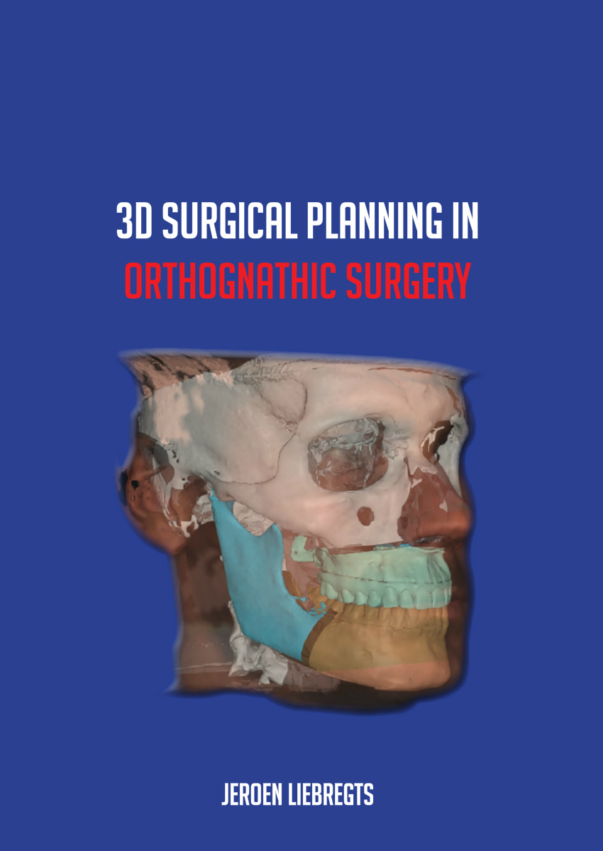
3D Surgical Planning in Orthognathic Surgery
Summary
In the past decades, numerous technological advances have had their influence on modern medicine. In orthognathic surgery, evolution in digital imaging technology has brought us the capability to visualize soft tissue, bony tissue, and dentition of a patient’s head in 3D. With this ability come many advantages, most substantially regarding 3D preoperative planning and 3D postoperative evaluation. In this thesis, the main objective is to investigate how 3D planning in orthognathic surgery can optimize the surgical outcome and predictability. In Chapter 1, a general introduction to the clinical and technical fundamentals of contemporary 3D orthognathic planning is provided. In order to create a virtual 3D head model, bony structures, soft tissue, and dentition need to be visualized. Different 3D imaging techniques are being used and the information from separate imaging modalities are combined using image fusion algorithms in dedicated software programs. Cone-beam computed tomography (CBCT) serves as the basis of a 3D virtual head model, as it provides information about the bone structures and thickness of overlying soft tissue. Combined with stereophotogrammetry, a 3D color photo of the soft tissue, an intra-oral scan of the dentition, an augmented 3D virtual head can be rendered. Virtual osteotomies can be performed (Le Fort I, bilateral sagittal split-osteotomy (BSSO), genioplasty) upon this 3D augmented head model, in which all jaw segments can be moved in any desired direction. The soft tissue outcome of a virtual osteotomy can be simulated in real-time by using different biomechanical models.
In Chapter 2, the accuracy of the Mass Tensor Model (MTM) algorithm in Maxilim software ® (Medicim NV, Mechelen, Belgium) is assessed. Pre- and postoperative CBCT scans of 100 patients that were treated with a BSSO were registered on the anterior cranial base. 3D distance maps and 3D cephalometric analyses were used to calculate the differences between the soft tissue simulation and the actual postoperative soft tissue results. For the entire face, the mean absolute error was found to be 0.9±0.3 mm and the mean absolute 90th percentile was 1.9 mm. The chin area was the most predictable area, with a mean absolute error of 0.8±0.5 mm, whereas the lower lip area was the least predictable, with a mean absolute error of 1.2±0.5 mm. It is important to realize that the lip area may be the most difficult area to predict. The presence of orthodontic appliances, or absence thereof, and the difficulty in maintaining the same amount of tension in patients’ lips in pre- and postoperative CBCTs might have a role in the pre- and postoperative discrepancy.
In Chapter 3, a similar study was conducted on the accuracy of soft tissue prediction in bimaxillary osteotomies. Sixty patients who underwent bimaxillary surgery were enrolled in this study. Using a similar methodology, a mean absolute error of 0.81±0.22 mm was found for the face as a whole. The accuracy of the soft tissue simulation in the upper lip region was the highest (1.2 ± 0.6 mm), whereas the lower lip region was found to be the least predictable (1.4 ± 0.5 mm). In general, the accuracy of the soft tissue simulation decreased in patients when a larger surgical advancement was planned.
In Chapter 4, the influence of Le Fort I osteotomy on the width of the nose at the alar base was assessed in 60 patients that underwent bimaxillary surgery. Despite using different closing sutures, the VY closure and alar cinch technique, widening of the alar base width was perceived, with an absolute mean of 1.6 ± 1.1 mm. The 3D cephalometric analysis of the simulation versus the actual postoperative results showed a mean absolute error of 1.0 ± 0.9 mm. A lack of correlation between the simulation error of alar widening and maxillary advancement was found, which seems to indicate that the MTM model used in this study was unable to simulate the correct relationship between postoperative alar changes and maxillary displacement and is therefore unsuitable for soft tissue prediction of the nasal region.
In 2014, a new 3D photogrammetry-based automated method for quantifying variations in soft tissue facial profiling using principal component analysis (PCA) was developed in the department of oral and maxillofacial surgery at Radboudumc to evaluate surgical related facial soft tissue changes. PCA is applied to reduce complex data in simplified structured patterns and is a commonly used tool in modern data analysis. It is based on statistical simulation rather than biomechanical models. The model is presented in Chapter 5. The PCA model can automatically quantify the facial appearance of the chin region of dysgnathic patients compared to a large group of controls. In this way, the effects of orthognathic surgery on the soft tissue facial profile can be evaluated. The effect of BSSO advancement surgery on the facial profile of Class II patients in comparison to a control sample of the Dutch population with a Class I facial profile was assessed using PCA. 3D photographs of 25 female patients that underwent a BSSO and 70 female controls were acquired. The specific facial variations were defined as unique variations and the effect of each unique variation (UV) on the soft tissue facial profile was investigated. The UVs that described the variations in the retrusion of the mandible were used to distinguish patients with mandibular hypoplasia from the control population. A clockwise rotation of the mandible and a shortening of the lower part of the face were the most prominent differences between the two groups (UVN). A protrusion of the upper lip and a retrusion of the mandible were observed among the preoperative BSSO patients compared to the control group. Consequently, an over-accentuation of the labial-mental fold was present in the preoperative BSSO patient group compared to the control group. This morphological variation was defined as UV2. For all subjects the scores for UVN and UV2 were calculated, the effect of BSSO advancement surgery was evident. The postoperative group of BSSO patients had shifted towards the control group compared to the preoperative situation. However, the postoperative group did not overlap the control group completely, indicating that many BSSO patients maintained some characteristics of Class II facial profile despite having had the surgery.
In the past, cephalometric landmarks were used to evaluate the accurate transfer of 2D and 3D planned bony movements to the actual postoperative result. However, this method is based on identifying the same landmarks multiple times which leaves room for error. It therefore impedes a correct interpretation of the analysis and the actual difference between the preoperative (3D) planning and postoperative outcome. Chapter 6 presents the OrthoGnathicAnalyser (OGA), a novel tool to quantify the displacement of bony segments in orthognathic surgery. Pre- and postoperative CBCT scans of ten patients who underwent bimaxillary surgery were acquired. To calculate the skeletal discrepancies between the 3D planning and the actual surgical outcome, the jaws were segmented and superimposed upon the postoperative maxillary and mandibular segments using voxel-based registration. This innovative method of quantifying the surgical displacement of jaw segments in six degrees of freedom (sagittal, vertical, and transverse translations, and pitch, roll, and yaw) was highly reproducible (intraclass correlation coefficients > 0.97). By using the OGA, the skeletal discrepancies between 3D planning and the surgical outcome can be objectified, which eliminates the need to identify cephalometric landmarks multiple times.
Using the OGA, the effect of sequencing on the predictability of bimaxillary surgery could be objectively assessed. In Chapter 7, 116 patients undergoing bimaxillary surgery were appraised, where 58 patients were operated on with a maxilla-first approach, and 58 patients had the mandible surgical procedure before the maxillary surgical procedure. It was found that the achievability of the anterior displacement of the maxilla was significantly higher in the maxilla-first group (0.5±2.5 mm) than in the mandible-first group (2.0±1.9 mm, p<0.01). Chapter 8 evaluates the postoperative skeletal stability one year following bimaxillary surgery in the same study cohort between the maxilla-first and mandible-first approaches. No significant differences were found in relapse between the maxilla-first or mandible-first groups. The study showed that the mean sagittal, vertical, and transverse relapse was less than 1.8 mm, which is consistent with previous findings. Chapter 9 evaluates the effects of sequencing in bimaxillary osteotomies in the study population of chapters 7 and 8 pooled with similar data from the Oral and Maxillofacial Surgery Research Unit at the University of Southern Denmark in Odense. The main conclusions of the chapter were that the maxilla-first sequencing was generally centered closer around the planned reposition than the mandible-first, while the mandible-first approach resulted in a significantly better AP position of the maxilla when an impaction of the maxilla was planned. Surgical accuracy of the maxilla for CCW or CW rotation was not statistically significantly influenced by sequencing. This thesis investigates the additional value of 3D preoperative planning and the postoperative 3D evaluation of the achieved surgical results. With 3D simulation, orthognathic surgery has become more accurate and predictable. Throughout this thesis, it became more and more clear that 3D simulation has become indispensable in orthognathic surgery, and indeed has now become the gold standard.
See also these dissertations


Sam Neppelenbroek
Dysregulation of autoreactive B cell responses in autoimmune diseases
9 juli 2026
Universiteit Leiden
Open Ebook


Karlijn Doorenspleet
Improving North Sea biodiversity monitoring using novel molecular approaches
25 juni 2026
Wageningen University
Open EbookWe print for the following universities


